Category: Parkinson's Disease: Neuroimaging
Objective: The aim of this study was to quantitatively evaluate iron content in idiopathic rapid eye movement sleep behavior disorder patients using quantitative susceptibility mapping.
Background: It is assumed that as the prodromal stage of α-synucleinopathies, idiopathic rapid eye movement sleep behavior disorder patients may have emerged iron deposition in brain.
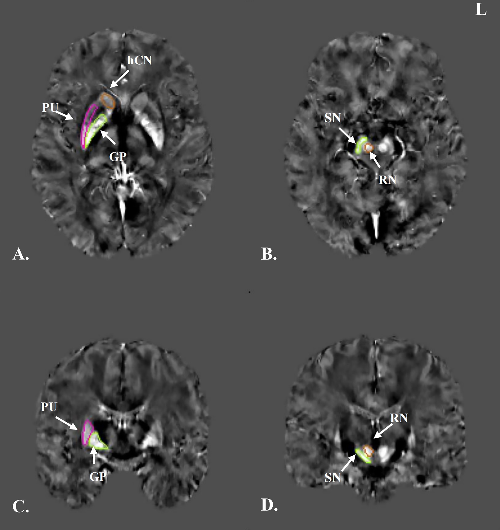
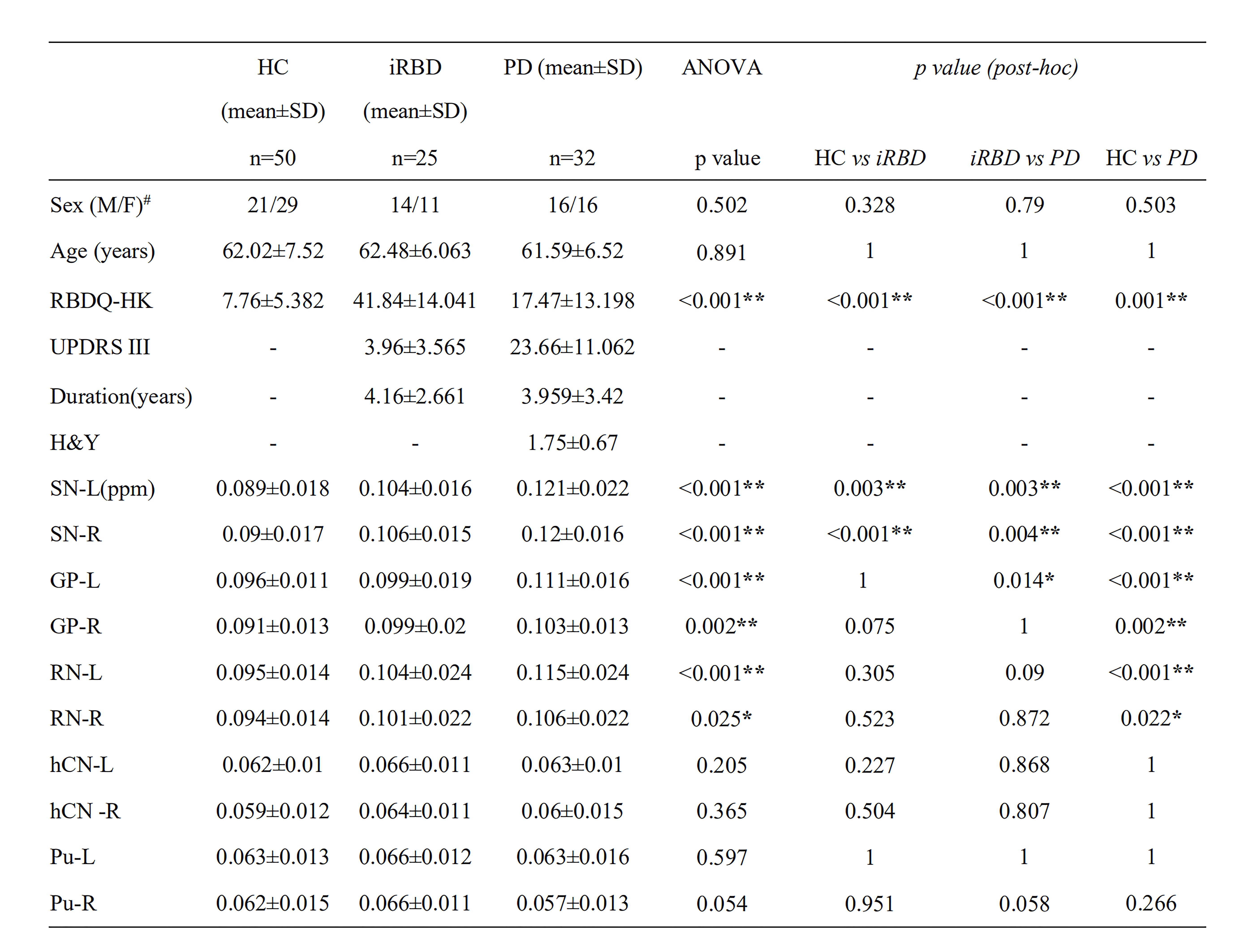
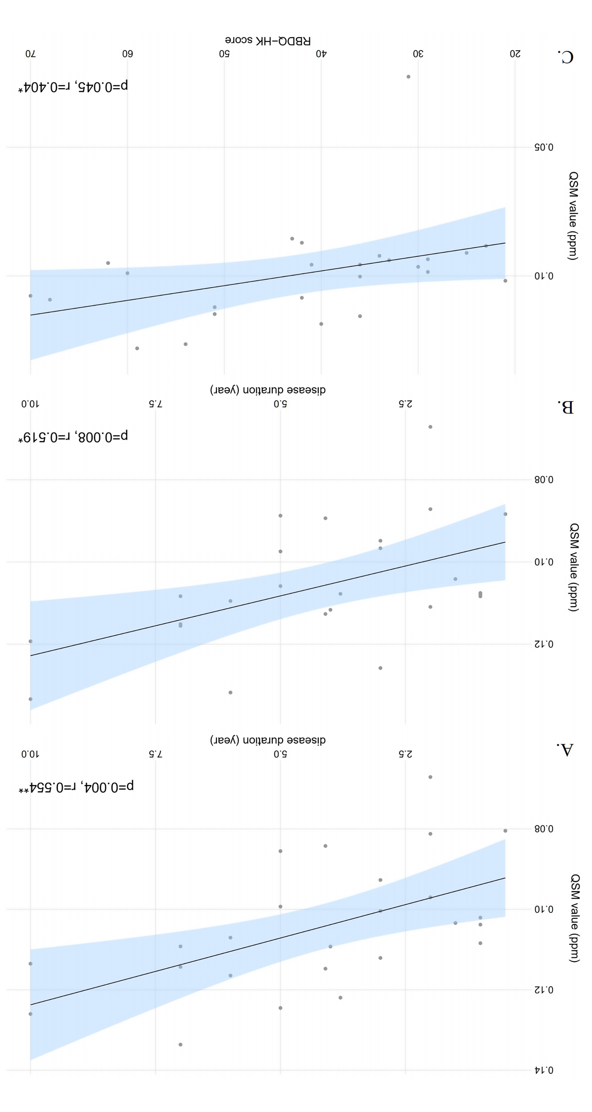
Method: Twenty-five idiopathic rapid eye movement sleep behavior disorder patients, 32 Parkinson’s disease patients, and 50 healthy controls underwent quantitative susceptibility mapping. We selected ten brain areas as regions-of-interest, including the substantia nigra, globus pallidus, red nucleus, the head of caudate nucleus and putamen bilaterally (Figure. 1). The mean susceptibility values within the ten brain areas were calculated and compared among groups (Table. 1). The relationships between the values and the clinical data of idiopathic rapid eye movement sleep behavior disorder and Parkinson’s disease were measured using correlation analysis (Figure. 2).
Results: Idiopathic rapid eye movement sleep behavior disorder patients had elevated iron in the bilateral substantia nigra compared to controls (post-hoc test, p < 0.005, Bonferroni corrected). Parkinson’s disease patients had increased iron in the bilateral substantia nigra, globus pallidus, and left red nucleus compared to healthy controls, and had elevated iron levels in the bilateral substantia nigra compared to idiopathic rapid eye movement sleep behavior disorder patients. Mapping values were positively correlated with disease duration in the left substantia nigra in the idiopathic rapid eye movement sleep behavior disorder patients. In addition, the mean susceptibility values had a tendency of positive correlation with the disease duration in the right substantia nigra, as well as had a tendency of positive relation with the rapid eye movement sleep behavior disorder questionnaire – Hong Kong scores in the right globus pallidus.
Conclusion: Quantitative susceptibility mapping can detect increased iron in the substantia nigra in idiopathic rapid eye movement sleep behavior disorder, which becomes more significant as the disorder progresses. This technique has the potential to be an early, objective neuroimaging marker for detecting α-synucleinopathies.
To cite this abstract in AMA style:
J. Sun, Z. Lai, J. Ma, L. Gao, M. Chen, J. Chen, J. Fang, Y. Fan, Y. Bao, D. Zhang, P. Chan, Q. Yang, C. Y, T. Wu, T. Ma. Quantitative Evaluation of Iron Content in Idiopathic Rapid Eye Movement Sleep Behavior Disorder [abstract]. Mov Disord. 2020; 35 (suppl 1). https://www.mdsabstracts.org/abstract/quantitative-evaluation-of-iron-content-in-idiopathic-rapid-eye-movement-sleep-behavior-disorder/. Accessed December 26, 2025.« Back to MDS Virtual Congress 2020
MDS Abstracts - https://www.mdsabstracts.org/abstract/quantitative-evaluation-of-iron-content-in-idiopathic-rapid-eye-movement-sleep-behavior-disorder/


