Category: Parkinson's Disease: Neuroimaging
Objective: 1. Identify the role of Cerebellum Basal Ganglia structural and functional connectivity changes in Healthy subjects Vs PD patients.
2. Establish Cerebellum Basal Ganglia interconnecting tracts in healthy and its changes in early and late stages of PD.
Background: The role of two-way interconnecting network between cerebellum and basal ganglia in the pathogenesis of PD in human subjects remains poorly understood [1]. We aim at identifying the role of Cerebellum Basal Ganglia structural and functional connectivity changes in the pathogenesis of PD. We hypothesize that, connectivity between basal ganglia and cerebellum (structural and functional) can be demonstrated by rs-fMRI and DTI in healthy volunteers and is altered in patients with PD in its early and late stages.
Method: We recruited 25 PD patients classified into ‘Early PD’ and ‘Late PD’ based on disease severity and duration along with a control group of 25 age and gender matched Healthy Subjects (HS). All the patients underwent the MRI session (Diffusion Tensor Imaging (DTI), functional MRI and structural 3D T1-w) and clinical assessment in their medication ‘ON’ and ‘OFF’ state whereas the HS group underwent single sessions. We located the seed regions in the Putamen (Dorsal/Causal), Thalamus, Cerebellar locomotor regions, Dentate Nucleus, Sub Thalamic Nucleus (STN) and Pontine Nuclei.
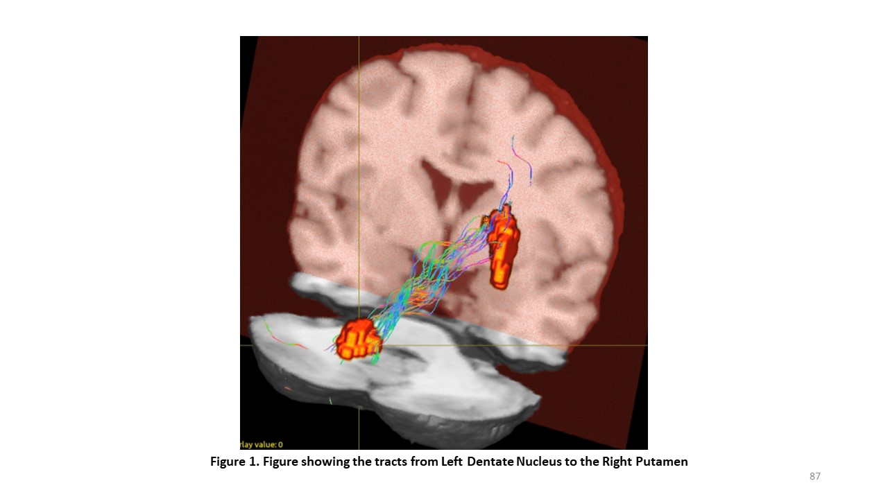
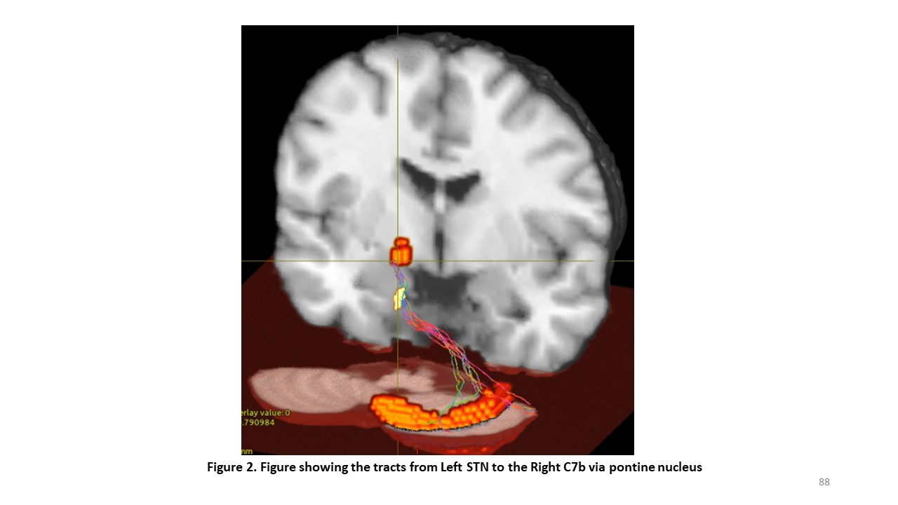
Results: In the HS group, DTI tractography demonstrated the two-way connection from the Dentate Nucleus to the Putamen via Thalamus [figure1]; STN to Cerebellum 7b via the Pontine Nucleus [figure2]. Reduced FC changes were observed between the Dorsal Putamen and Thalamus in the ‘Early PD’ group compared to HS group. In the ‘Late PD’ group, increased FC was observed between Cerebellum VIIIb and other cerebellar regions in comparison to HS. Also, the FC between Pontine Nuclei and cerebellar cortices were altered in the ‘Late PD’ group in comparison to HS. Increased FC was observed in ‘Early ON’ group between Putamen and Thalamus regions in comparison to the ‘Early OFF’ group.
Conclusion: The results suggest that, Cerebellum is involved at a later stage in the pathogenesis of PD and one of mechanisms through which Levodopa ameliorates symptoms in PD is reflected in the FC between thalamus and Dorsal Putamen. 5th Annual Conference of Movement Disorders Society of India, MDSICON2020, on February 2nd 2020.
References: [1] Bostan, A.C., Strick, P.L. The basal ganglia and the cerebellum: nodes in an integrated network. Nat Rev Neurosci 19, 338–350 (2018).
To cite this abstract in AMA style:
V. Radhakrishnan, S. Krishnan, B. Thomas, C. Gallea, C. Kesavadas, A. Kishore. Resting State Functional and Structural Connectivity Changes of Cerebellum Basal Ganglia Interconnecting Network in Parkinson’s Disease [abstract]. Mov Disord. 2020; 35 (suppl 1). https://www.mdsabstracts.org/abstract/resting-state-functional-and-structural-connectivity-changes-of-cerebellum-basal-ganglia-interconnecting-network-in-parkinsons-disease/. Accessed January 20, 2026.« Back to MDS Virtual Congress 2020
MDS Abstracts - https://www.mdsabstracts.org/abstract/resting-state-functional-and-structural-connectivity-changes-of-cerebellum-basal-ganglia-interconnecting-network-in-parkinsons-disease/


