Category: Parkinson's Disease: Neuroimaging
Objective: To explore the relationship between temporal resting-state functional magnetic resonance imaging (fMRI) properties and ON/OFF Levodopa motor performance scores in patients with Parkinson’s disease (PD).
Background: The mechanisms underlying deep brain stimulation (DBS) therapy are not fully understood. This has challenged individualized treatment strategies as well as prediction of therapeutic benefit. Evaluation of individual functional anatomy derived from rsfMRI holds great promise for knowledge advancement in this area.1 Compared to its spatial properties, the temporal properties of rsfMRI have been less explored,2 despite encoding multiple dimensions of information that may support more direct mapping of brain activity to clinical phenotypes.
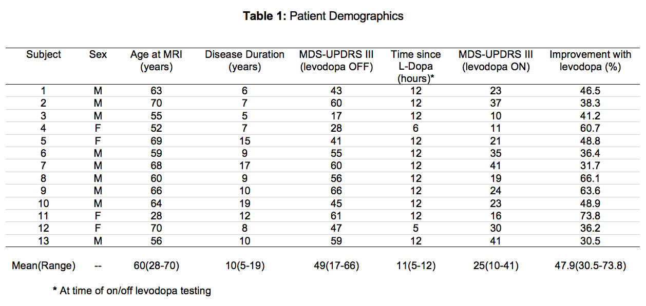
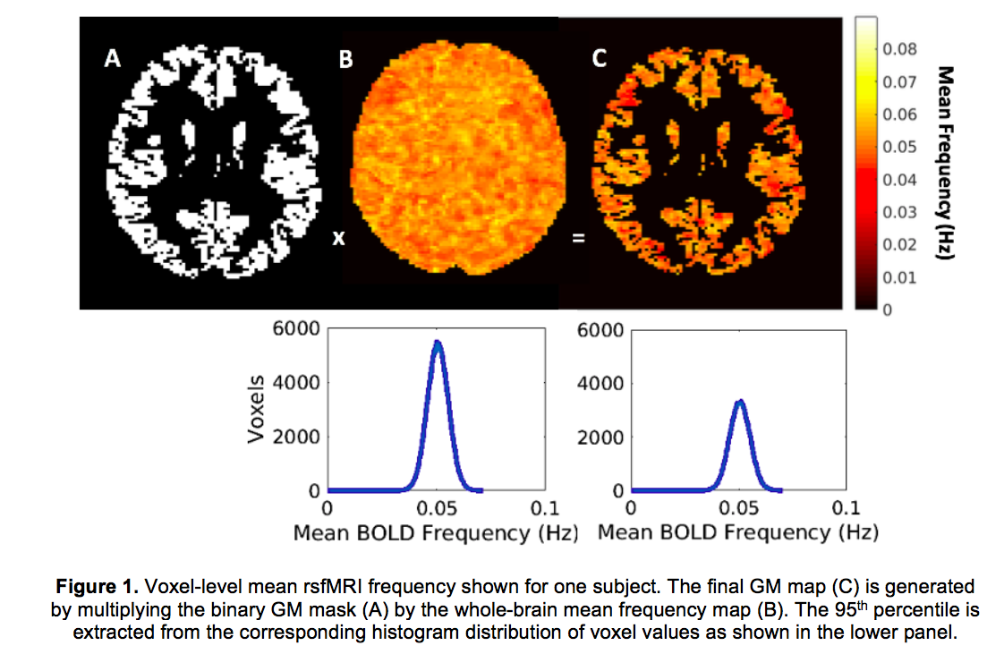
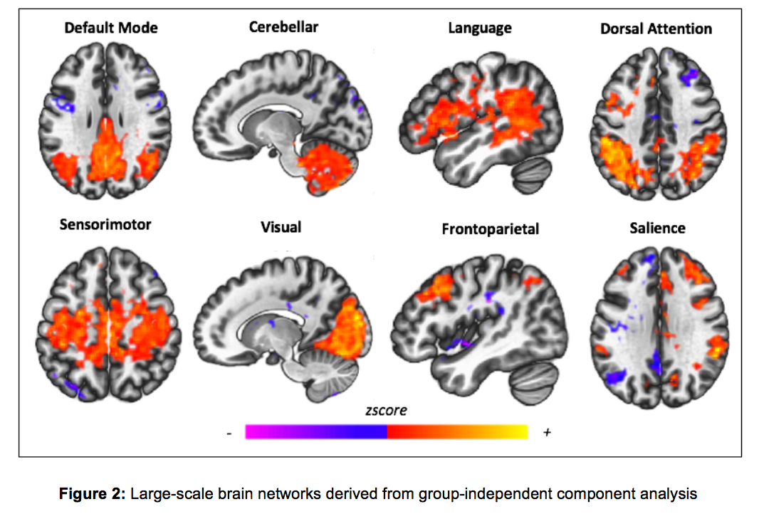
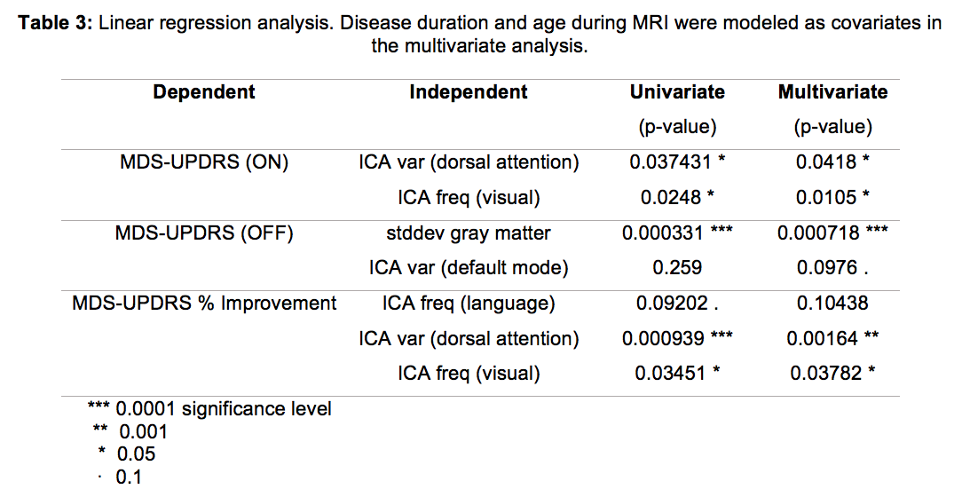
Method: Data for this study were retrospectively collected from 13 patients undergoing DBS implantation surgery for PD [table 1]. All MRIs were performed on a 3T GE system and included acquisition of axial T1-weighted images of brain anatomy and rsfMRI time series data [table 2]. The mean frequency and standard deviation of the rsfMRI signal were calculated for each voxel in the gray matter [figure 1]. Group independent component analysis and back-reconstruction were used to extract eight large-scale brain networks [figure 2] and map the frequency and variance signatures of each network back into subject-space. Univariate and multivariate linear regression assessed the relationship between imaging metrics and preoperative baseline motor performance during the ON and OFF levodopa states. Motor performance scores were calculated according to part III of the Movement Disorder Society – Unified Parkinson’s Disease Rating Scale (MDS-UPDRS III). Disease duration and age at the time of MRI were included as covariates in the multivariate analyses.
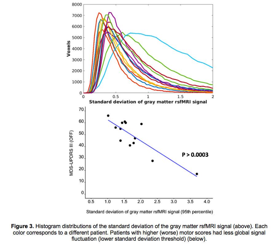
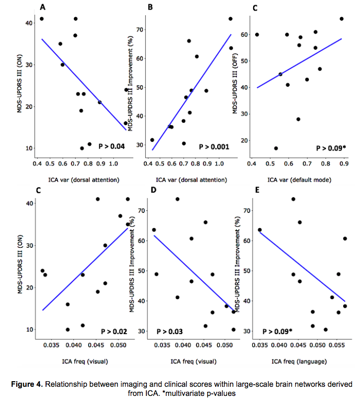
Results: Fluctuations in the gray matter rsfMRI signal were significantly associated with MDS-UPDRS III during the OFF levodopa state [figure 3]. In the dorsal attention network, rsfMRI signal fluctuations revealed the strongest association with percent MDS-UPDRS III improvement [figure 4; table 3].
Conclusion: Reduced rsfMRI signal fluctuations are associated with greater symptom severity and lower percent MDS-UPDRS improvement with levodopa. Thus, rsfMRI can effectively capture abnormal temporal patterns of rest activity and differentiate clinical phenotypes of PD.
References: 1. Boutet A, Rashid T, Hancu I, et al. Functional MRI Safety and Artifacts during Deep Brain Stimulation: Experience in 102 Patients. 2019. Neuroradiology. 293:174-183. 2. Venkatesh M, Jaja J, Pessoa L. Brain dynamics and temporal trajectories during task and naturalistic processing. 2019. NeuroImage. 186:410-423.
To cite this abstract in AMA style:
M. Morrison, A. Lee, S. Wang, A. Martin, I. Bledsoe, J. Ostrem, P. Larson, P. Starr, D. Wang. Temporal properties of resting-state functional MRI are associated with ON/OFF levodopa MDS-UPDRS III scores in patients with Parkinson’s disease [abstract]. Mov Disord. 2020; 35 (suppl 1). https://www.mdsabstracts.org/abstract/temporal-properties-of-resting-state-functional-mri-are-associated-with-on-off-levodopa-mds-updrs-iii-scores-in-patients-with-parkinsons-disease/. Accessed December 25, 2025.« Back to MDS Virtual Congress 2020
MDS Abstracts - https://www.mdsabstracts.org/abstract/temporal-properties-of-resting-state-functional-mri-are-associated-with-on-off-levodopa-mds-updrs-iii-scores-in-patients-with-parkinsons-disease/

