Category: Parkinson's Disease: Neuroimaging
Objective: Development of reliable imaging biomarkers of dopaminergic cell neurodegeneration is crucial to facilitate therapeutic drug trials in Parkinson’s disease (PD). Neuromelanin-sensitive (NM) MRI techniques have been efficient in identifying neurodegeneration in the substantia nigra pars compacta (SNc). We assessed SNc damage in early PD patients and evaluated the diagnostic performance and accuracy using Artificial Intelligence (AI)-based SNc segmentation method.
Background: PD is characterized by progressive loss of dopaminergic neurons in the SNc, which contains NM pigment, resulting in high T1-weighted signal on MRI [1]. NM signal diminishes early in the SNc of PD [2,3]. NM-MRI could be used to monitor disease progression and response to therapies, which requires characterizing the NM signal variations by investigating the SNc.
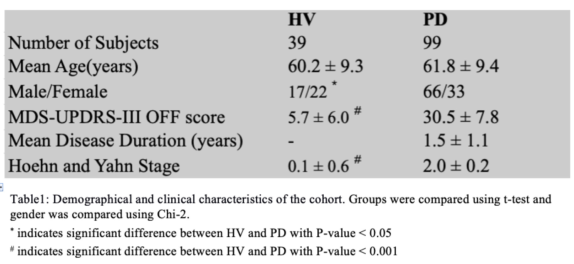
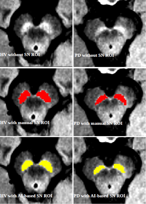
Method: We prospectively studied early PD cohort (Iceberg study) and age-matched healthy volunteers (HV) [table1]. Subjects were scanned at 3T MRI (PRISMA) using 3D T1-w and NM-sensitive images. We employed AI tools based on U-Net Convolutional Neural Network (CNN) architecture of deep learning for fully automated segmentation of SNc regions of interest (ROI) [4], which were validated using manually delineated SN ROI segmented by two expert raters [2,3] [figure1]. SNc Volume (Vol), corrected volume using total intracranial volume (Cvol), Signal-to-Noise-Ratio (SNR) and Contrast-to-Noise-Ratio (CNR) were computed. One-way GLM-ANOVA was conducted while adjusting for age and sex.
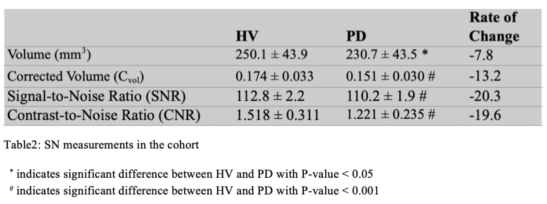
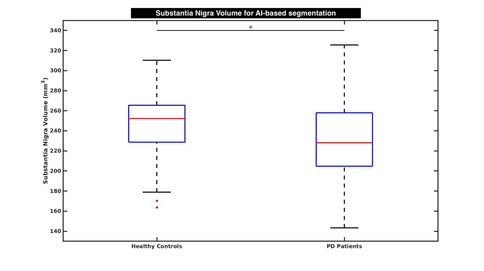
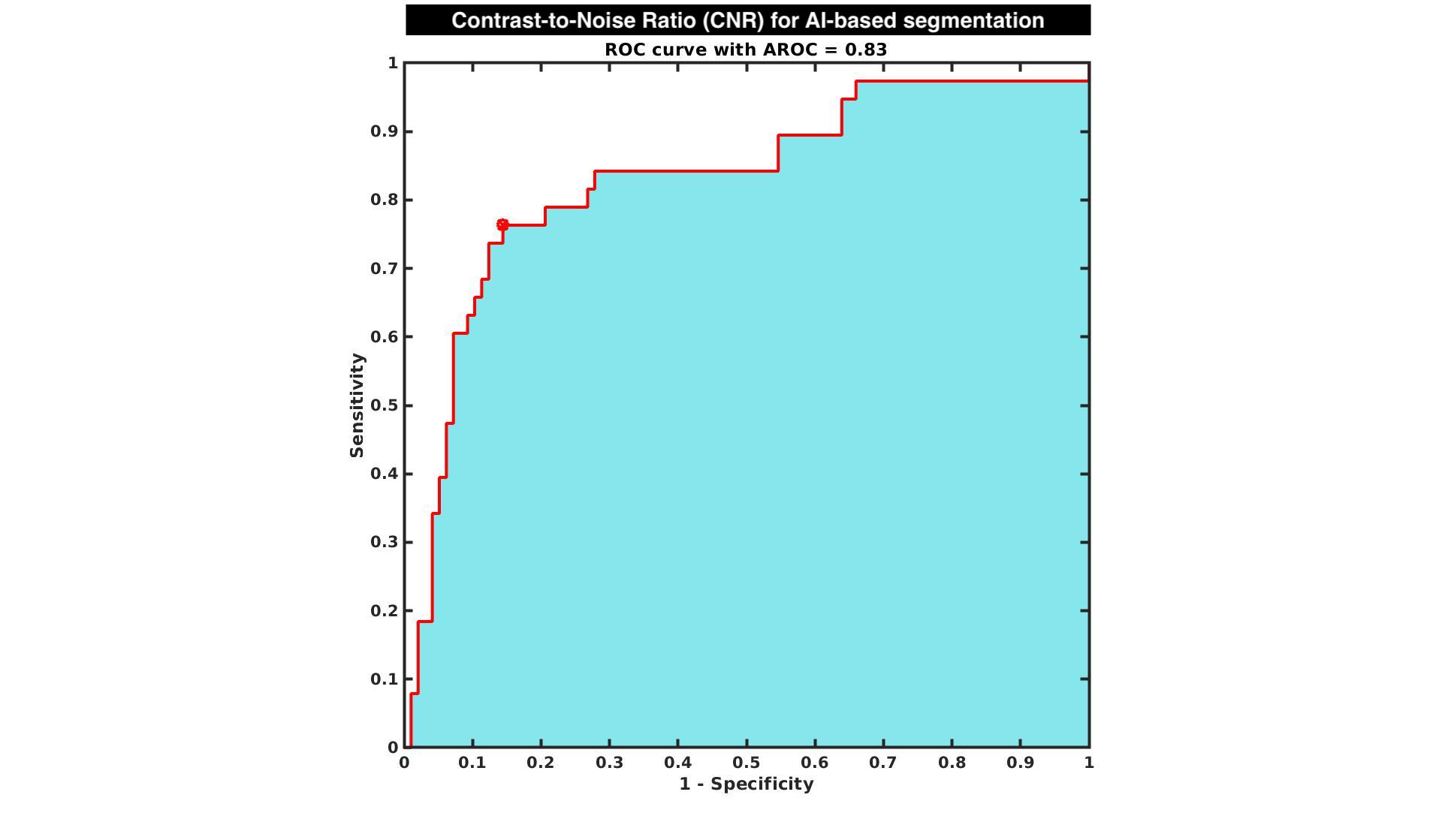
Results: Using AI-based segmentation method, PD patients demonstrated a highly significant effect for all SNc measurements with a reduction of 7.8% in Vol [figure1], 13.2% in Cvol, 20.3% in SNR and 19.6% in CNR relative to HV [table2] and ROC analysis provided area under the curve of 0.63 for Volume, 0.71 for Cvol, 0.80 for SNR and 0.83 for CNR [figure2] compared to manual of 0.71 for Vol, 0.75 for Cvol, 0.80 for SNR and 0.82 for CNR. Dice coefficient between AI-based and manual SNc ROI was 0.72 ± 0.08.
Conclusion: The proposed fully automated AI-based segmentation method showed comparable diagnostic performance with manual method which can possibly help us better understand the abnormalities in SNc. NM imaging might allow a direct non rater-dependent noninvasive evaluation of progression of SN cellular loss in PD and potential target for disease modification biomarker in drug trials.
References: 1. Sulzer, David, et al. “Neuromelanin detection by magnetic resonance imaging (MRI) and its promise as a biomarker for Parkinson’s disease.” NPJ Parkinson’s disease 4.1 (2018): 1-13. 2. Pyatigorskaya, Nadya, et al. “Magnetic resonance imaging biomarkers to assess substantia nigra damage in idiopathic rapid eye movement sleep behavior disorder.” Sleep 40.11 (2017): zsx149. 3. Pyatigorskaya, N., et al. “Comparative study of MRI biomarkers in the substantia nigra to discriminate idiopathic Parkinson disease.” American Journal of Neuroradiology 39.8 (2018): 1460-1467. 4. Ronneberger, Olaf, Philipp Fischer, and Thomas Brox. “U-net: Convolutional networks for biomedical image segmentation.” International Conference on Medical image computing and computer-assisted intervention. Springer, Cham, 2015. 5. Le Berre, Alice, et al. “Convolutional neural network-based segmentation can help in assessing the substantia nigra in neuromelanin MRI.” Neuroradiology 61.12 (2019): 1387-1395. 6. Shinde, Sumeet, et al. “Predictive markers for Parkinson’s disease using deep neural nets on neuromelanin sensitive MRI.” Neuroimage: Clinical 22 (2019): 101748.
To cite this abstract in AMA style:
R. Gaurav, R. Valabregue, L. Yahia-Cherif, N. Pyatigorskaya, G. Mangone, E. Biondetti, C. Ewenczyk, M. Hutchison, J.C Corvol, M. Vidailhet, S. Lehéricy. Investigating Substantia Nigra Damage using Fully Automated Segmentation of Neuromelanin MRI in Early Parkinson’s Disease [abstract]. Mov Disord. 2020; 35 (suppl 1). https://www.mdsabstracts.org/abstract/investigating-substantia-nigra-damage-using-fully-automated-segmentation-of-neuromelanin-mri-in-early-parkinsons-disease/. Accessed February 28, 2026.« Back to MDS Virtual Congress 2020
MDS Abstracts - https://www.mdsabstracts.org/abstract/investigating-substantia-nigra-damage-using-fully-automated-segmentation-of-neuromelanin-mri-in-early-parkinsons-disease/

