Session Information
Date: Wednesday, September 25, 2019
Session Title: Neuroimaging
Session Time: 1:15pm-2:45pm
Location: Les Muses Terrace, Level 3
Objective: We tested whether patients with Parkinson’s disease (PD) can increase the functional connectivity (FC) between the right insula (rIns) and dorsomedial frontal cortex (dmFC) using motor imagery guided by functional MRI (fMRI)-based neurofeedback (NF).
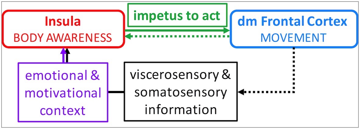
Background: A major cause of morbidity in PD is difficulty sustaining a steady motor performance. Intentional action is internally motivated and relies on body awareness. The rIns processes signals from the body and creates the impetus to act via its interaction with the dmFC. It then evaluates the outcome of the movements to reinforce adaptive movements (Fig1). Activation in both brain structures is diminished in patients with PD. NF may help them increase the FC between the two which in turn might lead to improvement in motor performance.
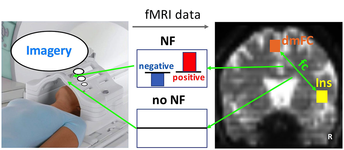
Method: 12 PD subjects (3 females, age: 64 ± 8.1 years) with mild bilateral disease were randomized to experimental motor imagery (MI, n=6) and control visual imagery (VI, n=6) groups. FMRI data were collected from all subjects in “on”-dopaminergic state in a 3T Siemens scanner. In the scanner, MI group practiced kinesthetic MI of complex body movements and received 10-12 NF training sessions on two days; VI group practiced VI of static scenes (e.g., lake, mountain) and did not receive NF training. The correlations between each subject’s rIns and dmFC activity patterns (i.e., FC) were calculated during 40-s imagery task blocks (x5). This FC was presented to the MI group as NF during training (Fig2). Both groups also practiced their respective imagery tasks at home for 15 min daily for 4 weeks. The first scan and last scan 4 weeks later were performed without NF in both groups and the difference in FC between them was used as a measure of learning success (two-way ANOVA).
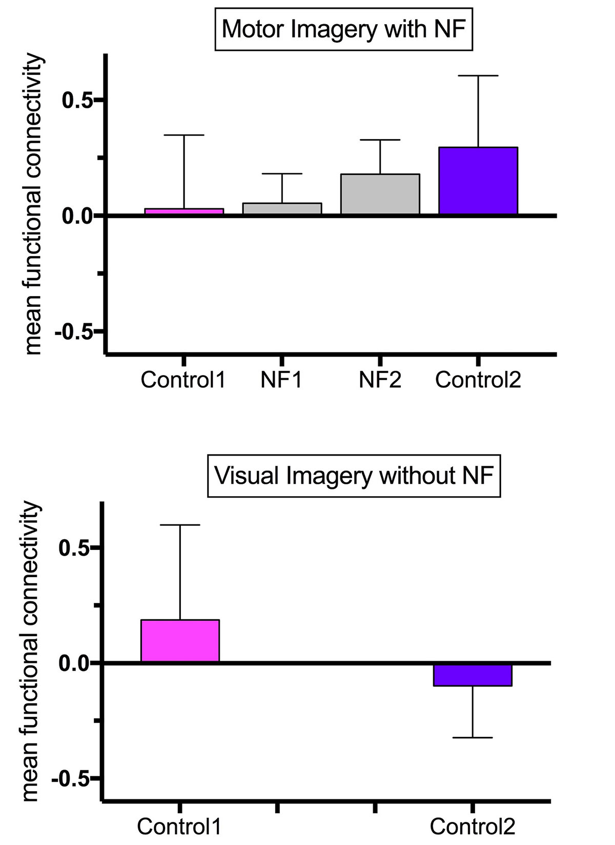
Results: There was a significant group-by-session interaction (p = 0.049) suggesting that the rIns-dmFC FC was significantly higher in the MI compared with the VI group as a result of training (Fig3). Frustration with imagined movements generated negative NF, whereas a feeling of ease and accomplishment generated positive NF. The most frequently reported MI themes were walking, gym exercises, and activities of daily living with a purpose to improve performance and focus.
Conclusion: PD patients can increase the FC between the rIns-dmFC. This increase is specific to the type of training, namely, NF-guided MI. Whether this increase predicts real world clinical outcomes needs to be studied further.
To cite this abstract in AMA style:
S. Tinaz, M. Elfil, J. Lemere, S. Basu, R. Sinha, D. Scheinost, M. Hampson. Neurofeedback-guided motor imagery in Parkinson’s disease [abstract]. Mov Disord. 2019; 34 (suppl 2). https://www.mdsabstracts.org/abstract/neurofeedback-guided-motor-imagery-in-parkinsons-disease/. Accessed April 2, 2025.« Back to 2019 International Congress
MDS Abstracts - https://www.mdsabstracts.org/abstract/neurofeedback-guided-motor-imagery-in-parkinsons-disease/


