Session Information
Date: Wednesday, September 25, 2019
Session Title: Cognition and Cognitive Disorders
Session Time: 1:15pm-2:45pm
Location: Agora 3 East, Level 3
Objective: To investigate the effects of levodopa on memory load and distractor resistance during visuospatial working memory (WM) in Parkinson’s disease (PD).
Background: Visuospatial WM deficits are one of the earliest cognitive changes in PD.1 Relative to verbal WM, individuals with PD have greater difficulty recalling features like the orientation, location or color of visual displays.2,3 Although few studies have examined the effects of levodopa on WM, the results are conflicting with two studies reporting beneficial effects4,5 and one study finding no effects.6 Visuospatial WM capacity is also affected by the ability to ignore distraction, which has not been well studied in PD.7 The effect of levodopa on distraction resistance in PD is less understood, with only one study finding beneficial effects.8 Altogether, there is a need to better understand the levodopa effects on both memory load and distraction resistance.
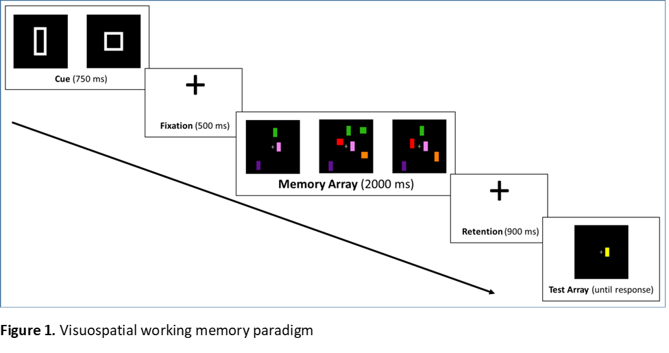
Method: 25 healthy controls (HC) and 28 PD patients without mild cognitive impairment (level 1 diagnostic criteria9) were recruited. The order of testing patients on and off levodopa was counterbalanced. To control for practice, the HC also performed the task twice in different sessions; half of HC data from the first session and half from the second session were analyzed. Figure 1 illustrates the paradigm. Memory array conditions were: 3 shapes no distractors, 3 shapes 3 distractors, and 5 shapes no distractors. The cue (rectangle or square) signaled which shape to attend to in the memory array. After the test array, individuals decided if the target was the same color and location as the shape in the memory array. Memory capacity (K) was analyzed (K= array size*(% hits – % false alarms). The effect of levodopa on K was tested separately for memory load (3 and 5 shapes without distraction) and distraction (3 shapes with and without distraction).
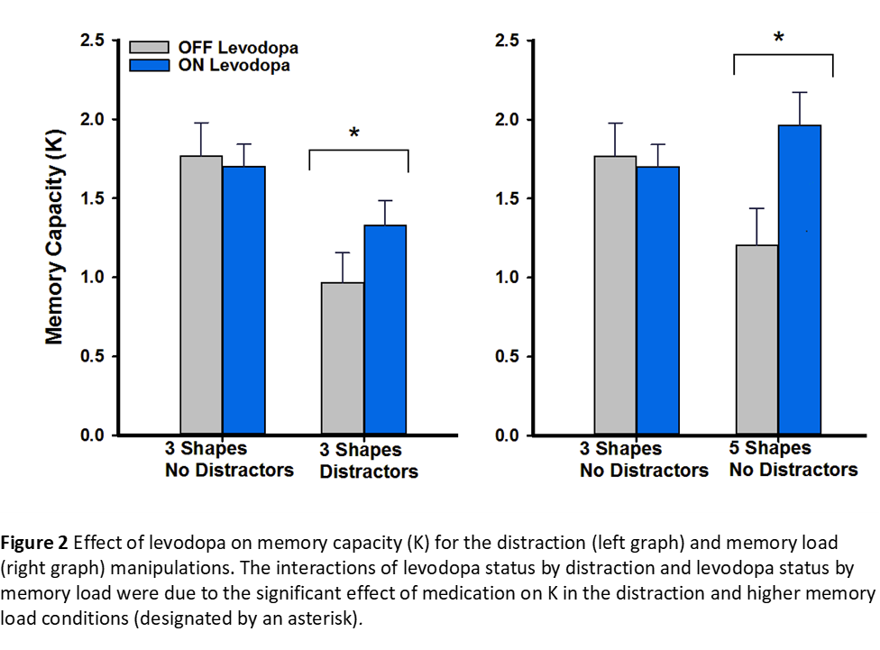
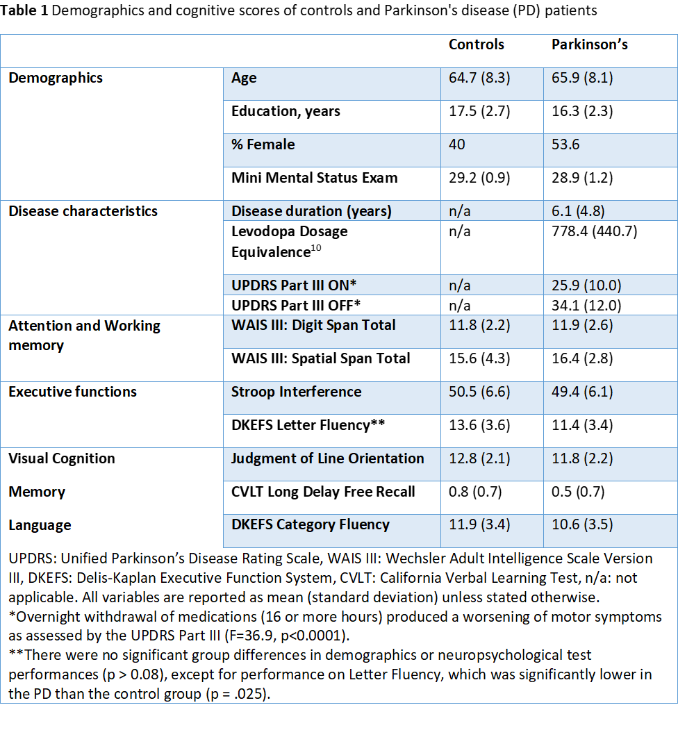
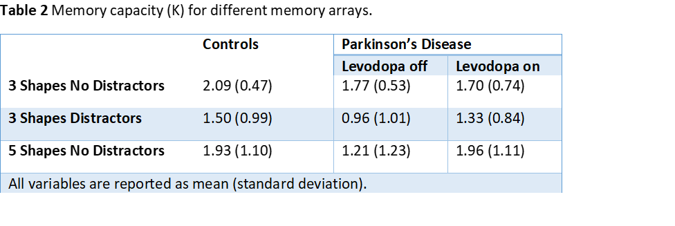
Results: Demographics and cognitive scores were similar between groups (Table 1) Medication Effects: Levodopa status interacted with distraction and memory load, improving K for the distraction (F=4.9, p=0.035) and larger memory load (F=19.4, p<0.0002) conditions (Figure 2). PD versus HC: Relative to HC, K was lower for distraction and higher memory load conditions in PD off, but not PD on levodopa (Table 2).
Conclusion: Levodopa restored visuospatial WM in PD, improving both memory capacity and resistance to distraction.
References: 1. Owen AM, Iddon JL, Hodges JR, Summers BA, Robbins TW. Spatial and non-spatial working memory at different stages of Parkinson’s disease. Neuropsychologia. 1997. doi:10.1016/S0028-3932(96)00101-7 2. Siegert RJ, Weatherall M, Taylor KD, Abernethy DA. A Meta-Analysis of Performance on Simple Span and More Complex Working Memory Tasks in Parkinson’s Disease. Neuropsychology. 2008. doi:10.1037/0894-4105.22.4.450 3. Zokaei N, Burnett Heyes S, Gorgoraptis N, Budhdeo S, Husain M. Working memory recall precision is a more sensitive index than span. J Neuropsychol. 2015;9(2):319-329. doi:10.1111/jnp.12052 4. Beato R, Levy R, Pillon B, et al. Working memory in Parkinson’s disease patients: Clinical features and response to levodopa. Arq Neuropsiquiatr. 2008. doi:10.1590/S0004-282X2008000200001 5. Costa A, Peppe A, Dell’Agnello G, et al. Dopaminergic Modulation of Visual-Spatial Working Memory in Parkinson’s Disease. Dement Geriatr Cogn Disord. 2003;15(2):55-66. doi:10.1159/000067968 6. Mollion H, Ventre-Dominey J, Dominey P., Broussolle E. Dissociable effects of dopaminergic therapy on spatial versus non-spatial working memory in Parkinson’s disease. Neuropsychologia. 2003;41(11):1442-1451. doi:10.1016/S0028-3932(03)00114-3 7. Lee EY, Cowan N, Vogel EK, Rolan T, Valle-Inclán F, Hackley SA. Visual working memory deficits in patients with Parkinson’s disease are due to both reduced storage capacity and impaired ability to filter out irrelevant information. Brain. 2010. doi:10.1093/brain/awq197 8. Fallon SJ, Mattiesing RM, Muhammed K, Manohar S, Husain M. Fractionating the neurocognitive mechanisms underlying working memory: Independent effects of dopamine and Parkinson’s disease. Cereb Cortex. 2017. doi:10.1093/cercor/bhx242 9. Litvan I, Goldman JG, Tröster AI, et al. Diagnostic criteria for mild cognitive impairment in Parkinson’s disease: Movement Disorder Society Task Force guidelines. Mov Disord. 2012;27(3):349-356. doi:10.1002/mds.24893 10. Tomlinson CL, Stowe R, Patel S, Rick C, Gray R, Clarke CE. Systematic review of levodopa dose equivalency reporting in Parkinson’s disease. Mov Disord. 2010;25(15):2649-2653. doi:10.1002/mds.23429
To cite this abstract in AMA style:
E. Bayram, I. Litvan, B. Wright, C. Grembowski, D. Harrington. Levodopa Effects on Visuospatial Working Memory in Parkinson’s Disease [abstract]. Mov Disord. 2019; 34 (suppl 2). https://www.mdsabstracts.org/abstract/levodopa-effects-on-visuospatial-working-memory-in-parkinsons-disease/. Accessed October 17, 2025.« Back to 2019 International Congress
MDS Abstracts - https://www.mdsabstracts.org/abstract/levodopa-effects-on-visuospatial-working-memory-in-parkinsons-disease/


