Session Information
Date: Tuesday, September 24, 2019
Session Title: Parkinsonisms and Parkinson-Plus
Session Time: 1:45pm-3:15pm
Location: Agora 3 West, Level 3
Objective: To evaluate the effect of electrode configuration and vertical electrode spacing on volume of tissue activated (VTA) in the globus pallidus internus (GPi) with directional deep brain stimulation (DBS)
Background: Directional DBS leads enable stimulation to be more precisely directed within targeted brain areas. Understanding the shape and size of the VTA for various monopolar or bipolar configurations can inform and expedite clinical programming for GPi-DBS.
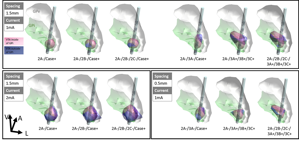
Method: A computational model was implemented in Sim4Life v4.0 with the multimodal image-based detailed anatomical (MIDA) model and a directional DBS lead with 1.5mm vertical electrode spacing placed with segmented contact 2 at the ventral-posterolateral GPi. DBS pulses of 1mA current with 60µs pulse width were used to produce VTAs. The following DBS contact configurations were tested: single-segment monopole (2A-/Case+), two-segment monopole (2A-/2B-/Case+ and 2A-/3A-/Case+), ring monopole (2A-/2B-/2C-/Case+), one-cathode, three-anode bipole (2A-/3A+/3B+/3C+), three-cathode, three-anode bipole (2A-/2B-/2C-/3A+/3B+/3C+). Additionally, certain vertical configurations were repeated for a directional DBS lead with 0.5mm vertical electrode spacing and also for 2mA current amplitude
Results: VTAs for monopolar configurations were spherically-shaped with the single-segment configuration generating the most directionality at the cathodic contact. When oriented correctly, the single-segment monopolar VTA was entirely encapsulated in the target region of the GPi, while two-segment and ring settings produced VTA that exceeded the GPi border inferiorly into the optic tract and medially into internal capsule. Bipolar configurations produced VTAs comparable to a monopolar setting at the level of the cathode and enlarged wedge-shaped VTAs at the level of the anode. Using a DBS lead with 0.5mm vertical electrode spacing further concentrated VTAs within the GPi.
Conclusion: We demonstrate that in GPi, single-segment monopolar configurations can improve targeting by forming more focused VTA at the cathode. Vertical bipolar settings can effectively enlarge the VTA at the anode without activating the side-effect areas surrounding the cathode. Using a DBS electrode with smaller vertical electrode spacing is beneficial for restricting VTAs to the GPi, while a larger vertical spacing can be used to capture more dorsal regions of the pallidum. [figure1]
To cite this abstract in AMA style:
S. Zhang, E. Pereira, N. Pouratian, A. Kent, B. Cheeran, L. Venkatesan, A. Schnitzler. Steering the Volume of Tissue Activated with Directional Deep Brain Stimulation Lead in the Globus Pallidus internus [abstract]. Mov Disord. 2019; 34 (suppl 2). https://www.mdsabstracts.org/abstract/steering-the-volume-of-tissue-activated-with-directional-deep-brain-stimulation-lead-in-the-globus-pallidus-internus/. Accessed February 10, 2026.« Back to 2019 International Congress
MDS Abstracts - https://www.mdsabstracts.org/abstract/steering-the-volume-of-tissue-activated-with-directional-deep-brain-stimulation-lead-in-the-globus-pallidus-internus/

