Session Information
Date: Tuesday, September 24, 2019
Session Title: Parkinsonisms and Parkinson-Plus
Session Time: 1:45pm-3:15pm
Location: Agora 3 West, Level 3
Objective: To explore the relative merits of automated MRI-based volumetric measures over visual assessment in the differential diagnosis of Parkinson’s disease (PD), parkinsonian variant of multiple system atrophy (MSA-P) and progressive supranuclear palsy (PSP).
Background: Parkinson’s disease (PD), parkinsonian variant of multiple system atrophy (MSA-P) and progressive supranuclear palsy (PSP) are common neurodegenerative forms of Parkinsonism. The differentiation among these disorders has been challenging.
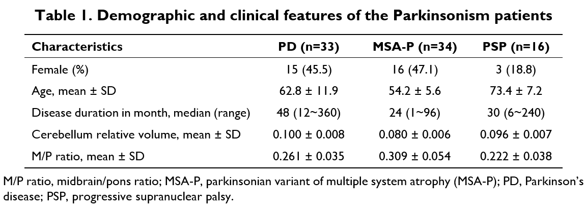
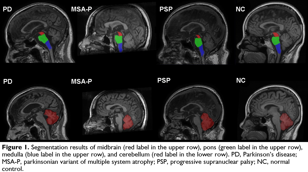
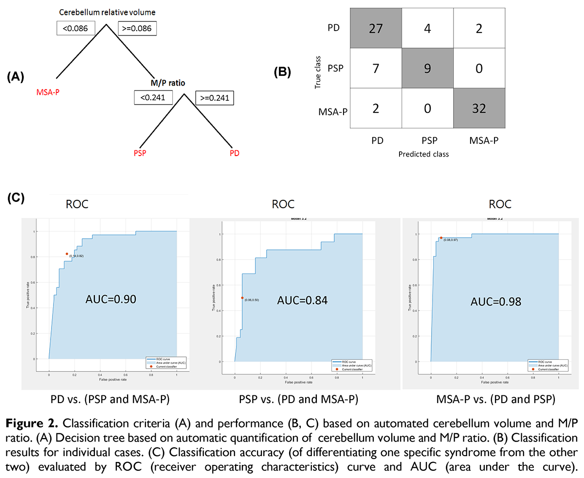
Method: We collected 3D T1-weighted MRI scans from 33 PD, 34 MSA-P and 16 PSP patients in this study [table1]. Differentiation of these disorders based on visual assessment was performed by an experienced neuroradiologist to detect the presence of “humming bird sign” as well as pontine and cerebellar atrophy. Differentiation with automated MRI biomarkers was achieved by calculating the cerebellum volume and midbrain to pons area ratio (M/P ratio), where optimal cutoff values of these two measures were determined. Segmentation and quantification of the cerebellum, midbrain and pons were performed with AccuBrain [figure1][1][2]. The performance of differentiational diagnosis by visual assessment and automated brain quantification was compared using decision tree-based classification as shown in [figure2](A) and [figure3](A).
Results: Using automated MRI-based quantitative biomarkers (i.e. cerebellum volume and M/P ratio) as predictive features, the multi-class classifier for differentiating PD, MSA-P and PSP achieved an overall accuracy of 81.9% (average AUC=0.907) [figure 2], while the overall classification accuracy of visual assessment was only 54.2% (average AUC=0.750) [figure 3].
Conclusion: Automated brain quantification presents a superior performance to visual assessment in the differentiation of PD, MSA-P and PSP. The automated MRI-based measurements of the cerebellum and brain stem may aid clinical differential diagnosis of the three parkinsonism disorders.
References: [1] Abrigo J, Shi L, Luo Y, Chen Q, Chu WCW, Mok VCT, et al. Standardization of hippocampus volumetry using automated brain structure volumetry tool for an initial alzheimer’s disease imaging biomarker. Acta Radiol. 2018:284185118795327 [2] Wang C, Zhao L, Luo Y, Liu J, Miao P, Wei S, et al. Structural covariance in subcortical stroke patients measured by automated mri-based volumetry. Neuroimage Clin. 2019;22:101682
To cite this abstract in AMA style:
H. You, Y. Luo, Y. Zhang, H. Wang, B. Hou, L. Shi, F. Feng. Automated Quantitative MRI Biomarkers in Differentiation of Progressive Supranuclear Palsy and Multiple System Atrophy from Parkinson Disease [abstract]. Mov Disord. 2019; 34 (suppl 2). https://www.mdsabstracts.org/abstract/automated-quantitative-mri-biomarkers-in-differentiation-of-progressive-supranuclear-palsy-and-multiple-system-atrophy-from-parkinson-disease/. Accessed April 2, 2025.« Back to 2019 International Congress
MDS Abstracts - https://www.mdsabstracts.org/abstract/automated-quantitative-mri-biomarkers-in-differentiation-of-progressive-supranuclear-palsy-and-multiple-system-atrophy-from-parkinson-disease/


