Session Information
Date: Monday, October 8, 2018
Session Title: Parkinson's Disease: Pathophysiology
Session Time: 1:15pm-2:45pm
Location: Hall 3FG
Objective: We hypothesize that cerebral hypoperfusion may influence the cognitive function of PD animal models. To investigate the effects of cerebral hypoperfusion and microangiopathy on Parkinson’s disease (PD) cognitive dysfunction in mice and the related mechanisms through the preparation of a PD mouse model of cerebral hypoperfusion.
Background: Previous report revealed that abnormal cerebral glucose metabolism and blood flow are related to the motor and cognitive symptoms underlying PD[1].
Methods: PD mice were prepared by intraperitoneal injection of MPTP and probenecid, and the mice were subsequently tested in the Morris water maze. The experimental mice were divided into 7 groups based on the cognitive results and bilateral common carotid artery stenosis (BCCAs) operation. After 28 days of stenosis, the mice in each group were subjected to pole climbing experiments. After behavioral testing, the brain tissue of each group was subjected to tyrosine hydroxylase (TH) staining, Nissl staining, Bielschowsky silver staining, TUNEL and Iba-1immunohistochemistry in each group. The levels of inflammatory cytokines in the plasma of each group were measured by chip-based liquid chromatography. Western blot was used to observe apoptosis and the expression of autophagy-related proteins in each group of mice.
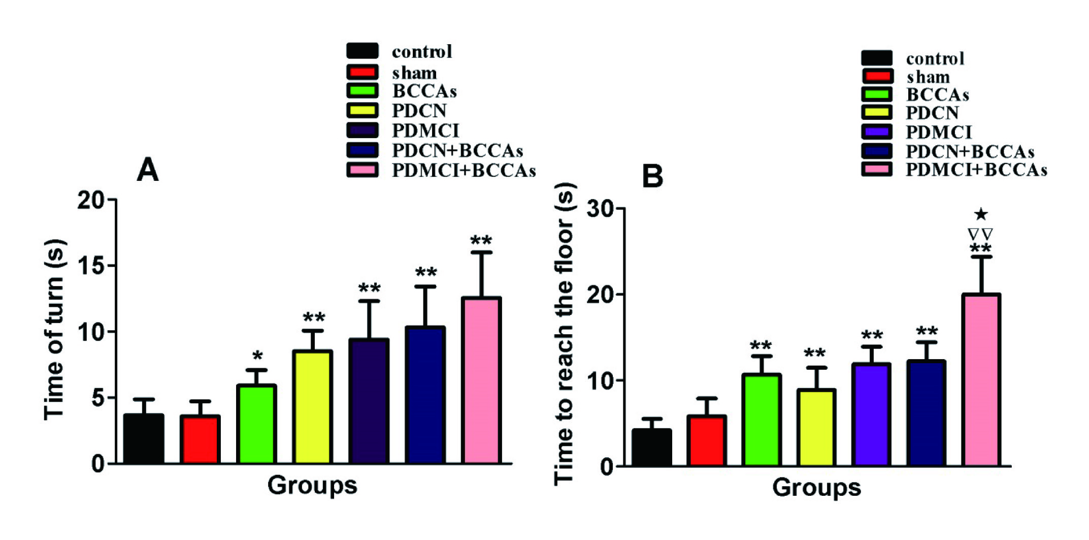
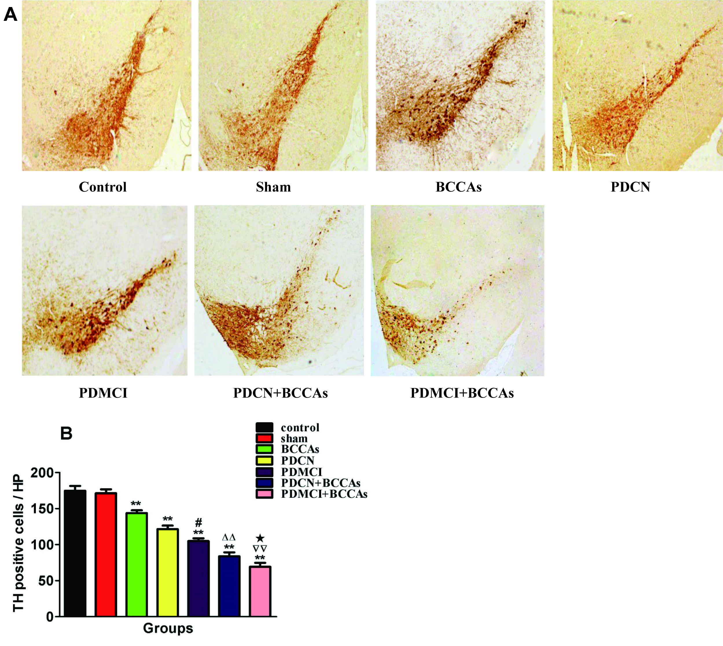
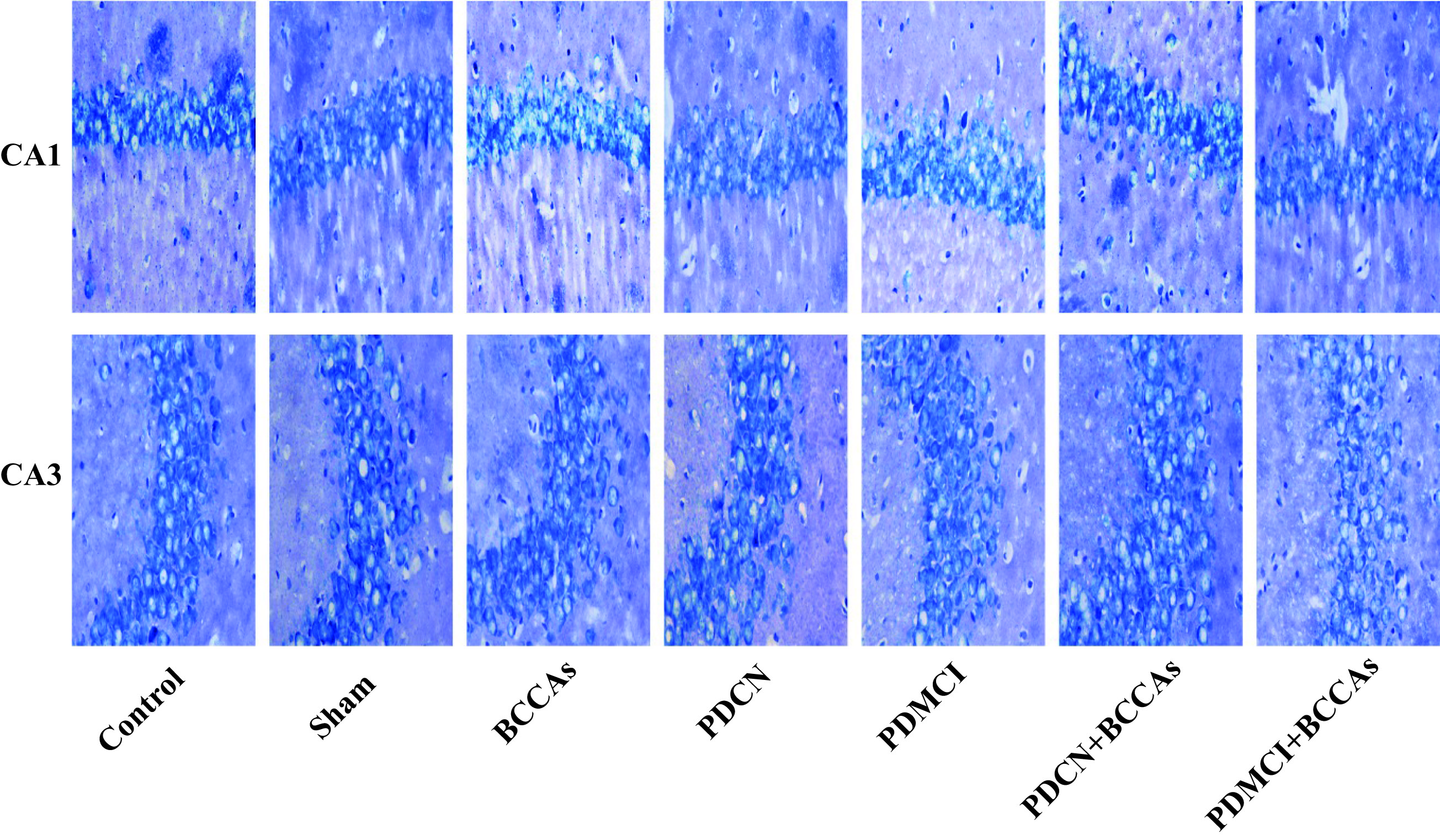
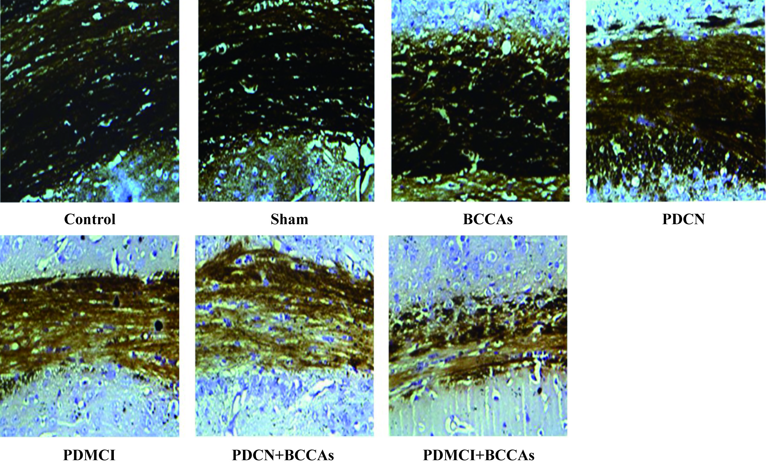
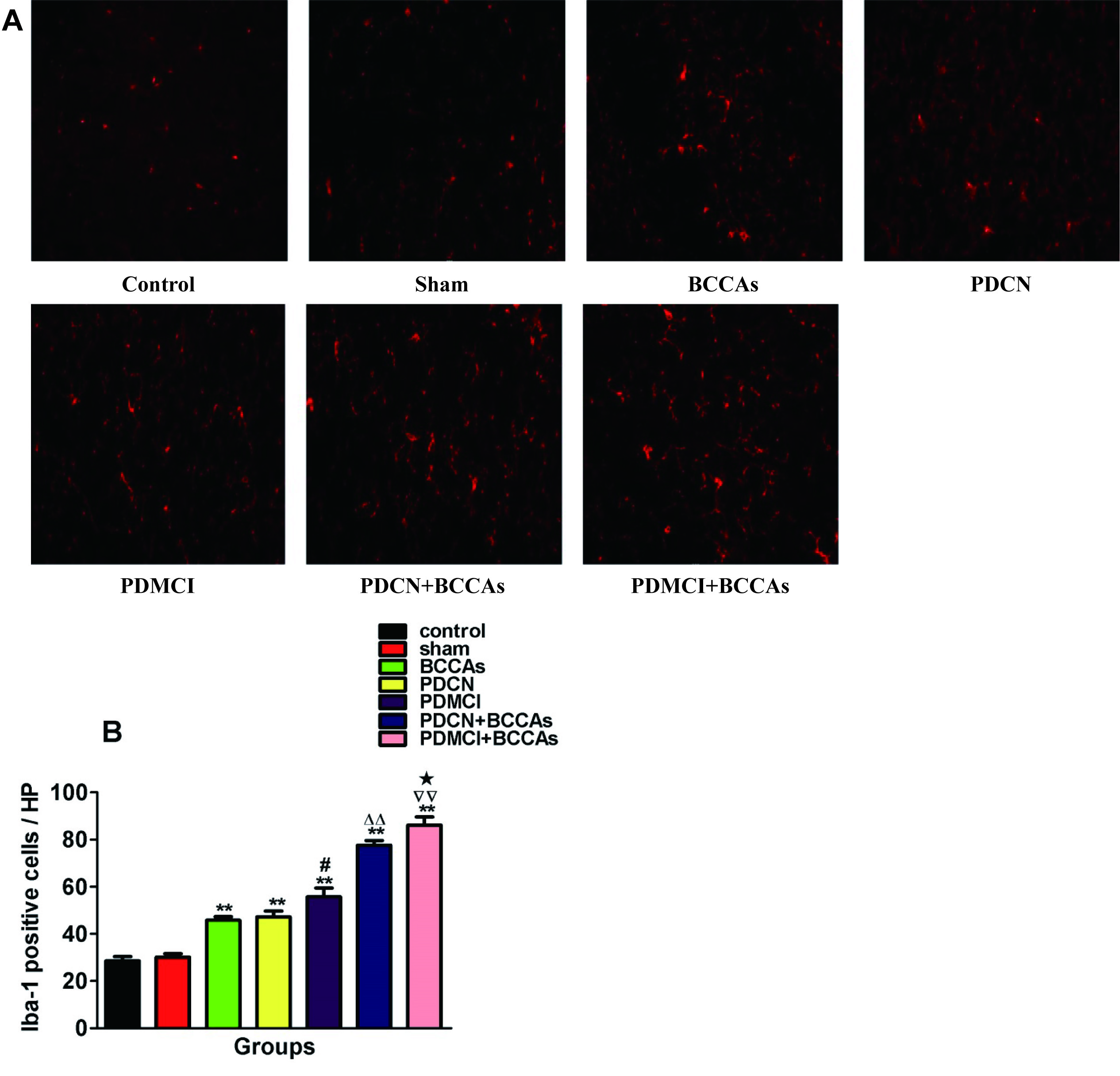
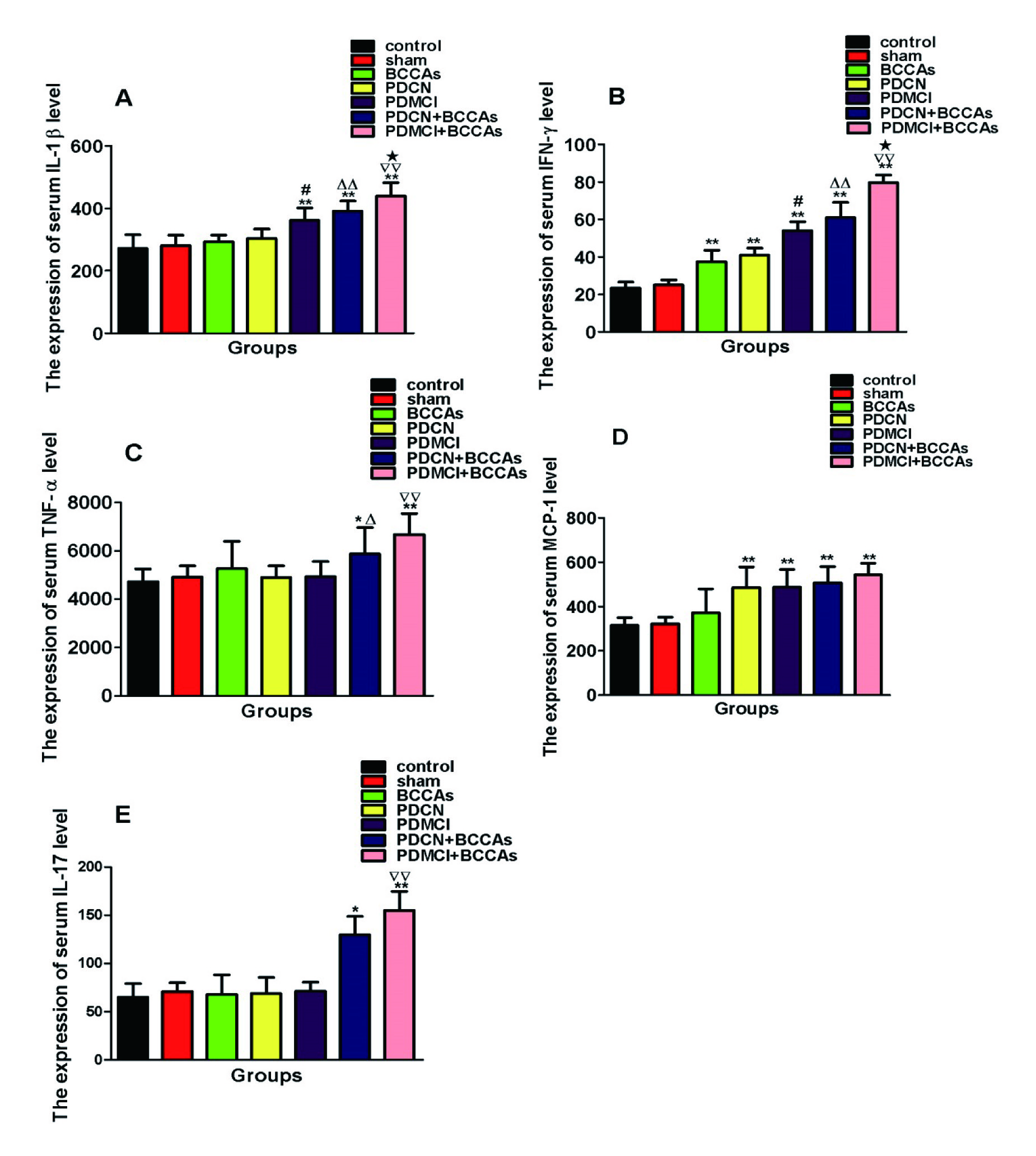
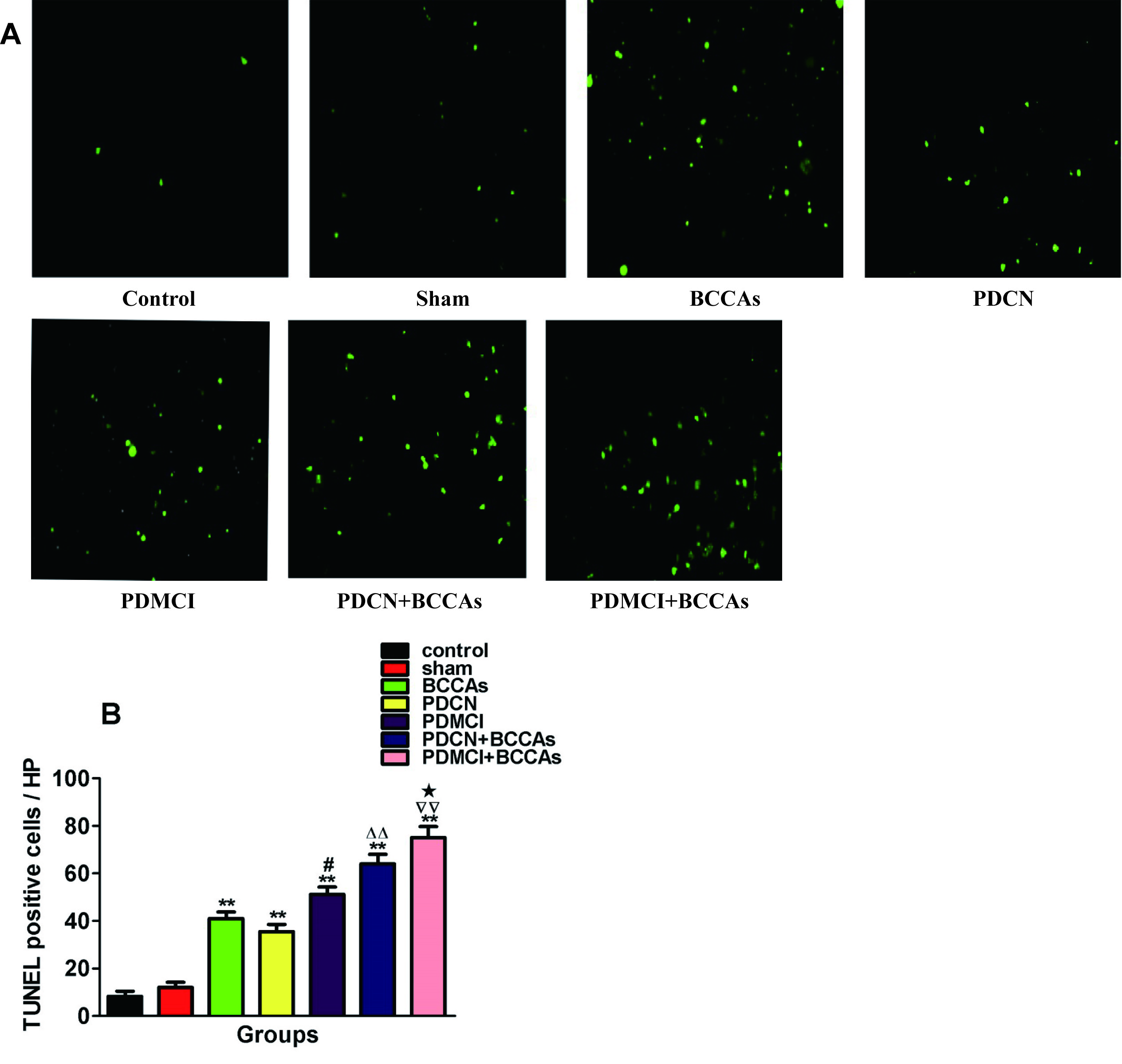
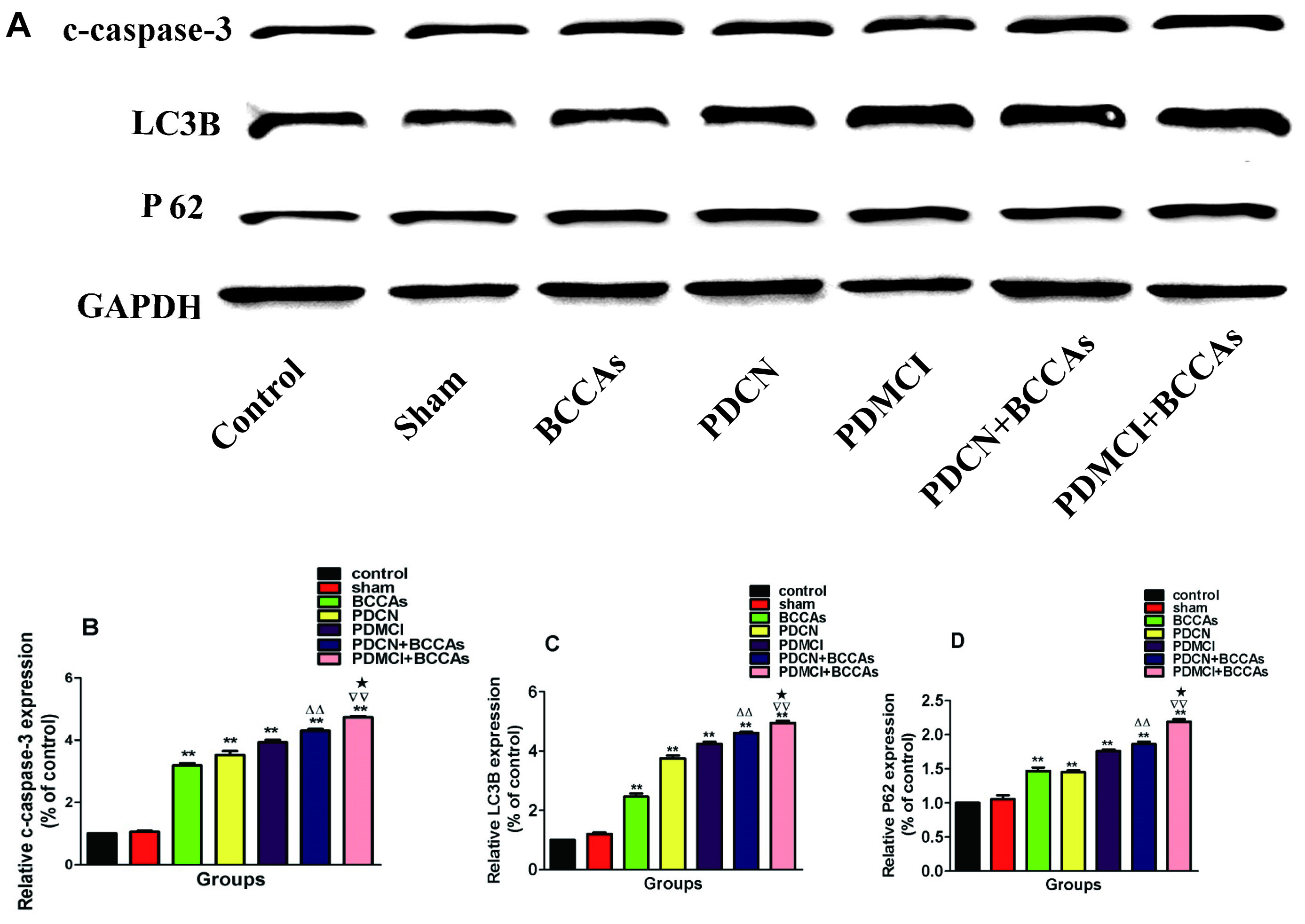
Results: (1) The pole-climbing experiment revealed that BCCAs could significantly prolong the climbing time of mice in the PDMCI group(Figure 1). (2) The immunohistochemistry results suggested that MPTP could reduce the number of TH-positive cells in the substantia nigra of mice, whereas BCCAs could significantly reduce dopamine (DA) neurons in the substantia nigra of PD mice(Figure 2). (3) Compared with the PDCN + BCCAs group, the PDMCI + BCCAs group exhibited white matter damage, significantly increased microglial activation (P < 0.01) and significantly increased levels of IL-1β and IFN-γ (P < 0.05)(Figure 5,6). (4) Nissl staining, TUNEL immunohistochemistry and Western blot revealed that MPTP injection alone or BCCAs alone could induce neuronal structural changes, reduce neuronal numbers, and increase neuronal apoptosis and that MPTP combined with hypoperfusion could promote the destruction of neuronal structures and neuronal apoptosis. In addition, we also found that the level of intracellular autophagy exhibited a compensatory increase(Figure 3,4,7,8).
Conclusions: Cerebral hypoperfusion can aggravate the impairment of cognitive function in PD mice. This finding may be related to hypoperfusion-mediated deterioration of neuroinflammation, aggravation of white matter damage, promotion of hippocampal neuron apoptosis and activation of autophagy in PD mice.
References: [1] Peng, S., D. Eidelberg, and Y. Ma, Brain network markers of abnormal cerebral glucose metabolism and blood flow in Parkinson’s disease. Neurosci Bull, 2014. 30(5): p. 823-37.
To cite this abstract in AMA style:
Y. Gao, H. Tang, K. Nie, R. Zhu, L. Gao, S. Feng, L. Wang, J. Zhao, Z. Huang, Y. Zhang, L. Wang. Microglial activation, white matter and hippocampal damage correlate with cognitive impairment in chronic cerebral hypoperfused and MPTP-lesioned mice [abstract]. Mov Disord. 2018; 33 (suppl 2). https://www.mdsabstracts.org/abstract/microglial-activation-white-matter-and-hippocampal-damage-correlate-with-cognitive-impairment-in-chronic-cerebral-hypoperfused-and-mptp-lesioned-mice/. Accessed November 8, 2025.« Back to 2018 International Congress
MDS Abstracts - https://www.mdsabstracts.org/abstract/microglial-activation-white-matter-and-hippocampal-damage-correlate-with-cognitive-impairment-in-chronic-cerebral-hypoperfused-and-mptp-lesioned-mice/

