Session Information
Date: Monday, October 8, 2018
Session Title: Parkinson's Disease: Neuroimaging And Neurophysiology
Session Time: 1:15pm-2:45pm
Location: Hall 3FG
Objective: To study the role of hippocampal subfield pathology in the genesis of psychotic symptoms in patients with Parkinson’s disease (PD) by comparing the volumes of several subfields of hippocampus in the MR images of a cohort of patients of PD (with and without psychosis) and controls.
Background: Psychosis, manifested through formed visual hallucinations or minor hallucinations, is a common non-motor symptom of PD. The pathogenesis of psychosis in PD remains unclear; however, is possibly linked to structural and functional alterations in the hippocampus. The literature on role of hippocampal pathology in the genesis of psychotic symptoms in PD is limited. To explore the role of hippocampus in psychosis, a detailed hippocampal subfield analysis was performed on PD patients with (PD-P) and without psychosis (PD-NP), and controls.
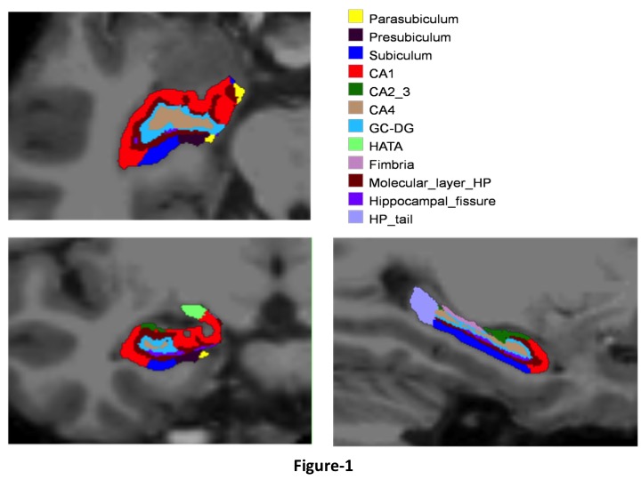
Methods: An automated subfield parcellation (using FreeSurfer 6.0) was performed on T1 MRI images of 69 subjects (PD-P: 28, PD-NP: 24, HC: 17). The volumes of 12 subfields on each side were estimated and analyzed between the three groups (Figure-1). Multiple comparisons were accounted for by employing false discovery rates with a significance level of 0.05. The volumes were also correlated to the neuropsychological tests assessing memory, visuo-spatial functions, and attention.
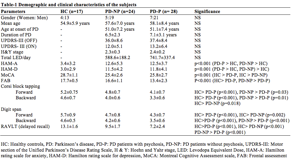
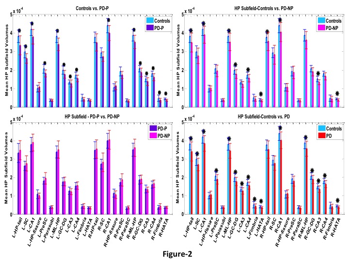
Results: The three groups were comparable in terms of age, gender, and education. Disease severity, stage of PD were comparable between the two PD-subgroups (Table-1). The two PD groups had similar Hippocampal subfield volumes were not significantly different between PD-P and PD-NP. Compared to controls, both the PD subgroups had lower volumes in bilateral CA1, hippocampus amygdala transition area, left molecular layer, granule cell-dentate gyrus, CA4, hippocampal tail, and right CA3. In addition, left subiculum and presubiculum, and right fimbria had significantly lower volumes in PD-P group compared to controls (figure-2). Significant positive correlations were observed between several subfields and cognitive scores, only in the PD-P group.
Conclusions: PD-P and PD-NP have no difference in the volumes of the hippocampal subfields. Our findings indicate a higher degeneration of specific hippocampal subfields in PD-P group compared to controls and these changes were correlated with the cognitive scores.
References: 1) Lenka A, Herath P, Christopher R, Pal PK (2017) Psychosis in Parkinson’s disease: From the soft signs to the hard science. J. Neurol. Sci. 379:169–176. 2) Yao N, Cheung C, Pang S, et al (2016) Multimodal MRI of the hippocampus in Parkinson’s disease with visual hallucinations. Brain Struct Funct 221:287–300. doi: 10.1007/s00429-014-0907-5.
To cite this abstract in AMA style:
A. Lenka, M. Ingalhalikar, A. Shah, J. Saini, S. Hegde, A. Shyam Sundar, R. Yadav, P. Pal. In vivo MRI signatures of hippocampal subfield pathology in patients with Parkinson’s disease and psychosis [abstract]. Mov Disord. 2018; 33 (suppl 2). https://www.mdsabstracts.org/abstract/in-vivo-mri-signatures-of-hippocampal-subfield-pathology-in-patients-with-parkinsons-disease-and-psychosis/. Accessed February 12, 2026.« Back to 2018 International Congress
MDS Abstracts - https://www.mdsabstracts.org/abstract/in-vivo-mri-signatures-of-hippocampal-subfield-pathology-in-patients-with-parkinsons-disease-and-psychosis/


