Session Information
Date: Monday, October 8, 2018
Session Title: Parkinson's Disease: Neuroimaging And Neurophysiology
Session Time: 1:15pm-2:45pm
Location: Hall 3FG
Objective: To explore the association between novel diffusion-derived structural imaging biomarkers and disease duration in Parkinson’s disease with freezing of gait (PD-FOG).
Background: Increasing free-water fraction in the posterior substantia nigra has been shown to correlate with disease progression in a large cohort of de-novo patients with Parkinson’s disease (PD)1. However, this correlation has not been investigated in patients with PD-FOG. Additionally, regions impaired in patients with PD-FOG may have differentiated free-water and a novel weighted fractional anisotropy (FAwt)2 measure was compared to patients with PD and healthy controls (HC). These novel imaging biomarkers may be associated with disease duration and severity in PD-FOG.
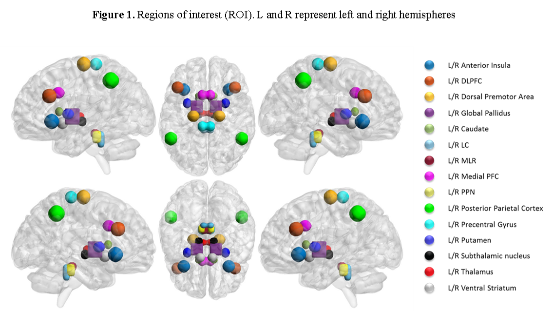
Methods: 8 HC, 7 PD-FOG, and 4 PD without FOG (PD-nFOG) were recruited for Center for Neurodegeneration and Translational Neuroscience. The group demographics are outlined [table 1]. All participants underwent structural magnetic resonance imaging using a 3T scanner. 15 bilateral regions of interest (ROIs) previously reported to be affected in PD-FOG were evaluated [Fig.1]. Conventional single tensor voxelwise measures of fractional anisotropy, mean diffusivity (MD), radial diffusivity (RD), and axial diffusivity (AxD), along with free-water corrected measures and FAwt were computed. These measures were compared between groups, and their association with symptom duration and severity was investigated.
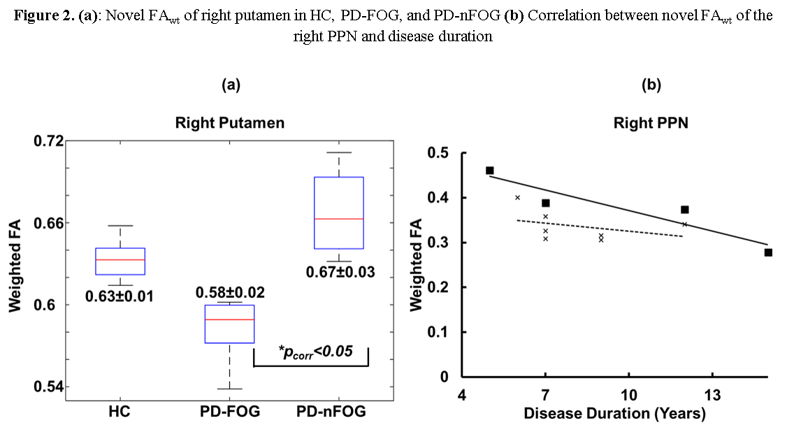
Results: Conventional single tensor measures along with free-water showed no group differences or association with symptom duration or severity. Only FAwt in the right putamen was significantly lower in PD-FOG compared to HC and PD-nFOG [Fig.2a]. Symptom duration was inversely correlated (p<0.05; [Fig.2b]) with FAwt of the right pedunculopontine nucleus (PPN) in patients with PD-nFOG. No significant correlations were found for PD-FOG.
Conclusions: Conventional single tensor measures along with free-water were not different in PD-FOG and did not have associations with symptom duration or severity. However, novel FAwt of right putamen was significantly lower in FOG even in this small sample. On the other hand, FAwt of the right PPN was not associated with symptom duration. Novel FAwt may be more sensitive in identifying structural breakdown in regions associated with PD-FOG. Data collected from a larger sample within this ongoing study will provide more reliable results.
References: 1 Burciu RG, Ofori E, Archer DB, et al. Progression marker of Parkinson’s disease: a 4-year multi-site imaging study. Brain 2017; 140: 2183–92. 2 Mishra V, Guo X, Delgado MR, Huang H. Toward tract-specific fractional anisotropy (TSFA) at crossing-fiber regions with clinical diffusion MRI. Magn Reson Med 2014; 0: 1–12.
To cite this abstract in AMA style:
B. Bluett, E. Bayram, S. Banks, Z. Mari, K. Sreenivasan, X. Zhuang, Z. Yang, C. Bird, D. Cordes, I. Litvan, V. Mishra. Exploring novel diffusion-derived biomarkers in Parkinson’s disease Freezing of Gait [abstract]. Mov Disord. 2018; 33 (suppl 2). https://www.mdsabstracts.org/abstract/exploring-novel-diffusion-derived-biomarkers-in-parkinsons-disease-freezing-of-gait/. Accessed February 5, 2026.« Back to 2018 International Congress
MDS Abstracts - https://www.mdsabstracts.org/abstract/exploring-novel-diffusion-derived-biomarkers-in-parkinsons-disease-freezing-of-gait/


