Session Information
Date: Monday, October 8, 2018
Session Title: Parkinson's Disease: Neuroimaging And Neurophysiology
Session Time: 1:15pm-2:45pm
Location: Hall 3FG
Objective: The cerebellum is involved in Parkinson’s disease (PD). This study was to investigate, using MRI and voxel-based morphometry (VBM), morphometric changes of cerebellum in PD with motor and non-motor subtypes.
Background: Parkinson’s disease (PD) is highly heterogeneous in clinical characteristics and outcomes. Subgroups have been defined both on the basis of motor and nonmotor features, which will require different disease-modifying and symptomatic treatments. As to motor features, Parkinson’s disease is sorted into tremor-dominant and postural instability gait disorder (PIGD) phenotypes. Furthermore, specific clinical descriptions of NMS-dominant phenotypes, particularly in the untreated PD model, are being increasingly described. Previous pathological and neuroimaging studies supported for fundamental biological differences between these different subtypes. More recent evidence has highlighted the role of the cerebellum. Up to date, the current results are still controversial and limited. In addition, there are few studies exploring the association between the cerebellum and the emotional disorders in PD, such as depressive and anxious symptoms.
Methods: 54 patients with idiopathic PD were classified into tremor-predominant-PD (PDT) (n=37) and akinetic/rigidity-predominant-PD (PDAR) (n=17). Moreover, they were classified as non-depressed PD (NDPD, n=41) and depressed PD (DPD, n=13), non-anxious PD (NAPD, n=38) and anxious PD (APD, n = 16), respectively. They were additionally compared at a group level with 39 normal controls (NC). We designed an analysis of covariance to examine cerebellar gray matter (GM) volume in different subgroups.
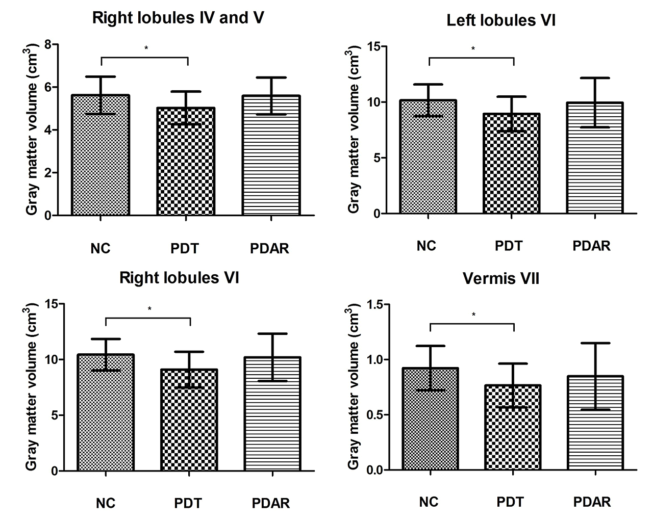
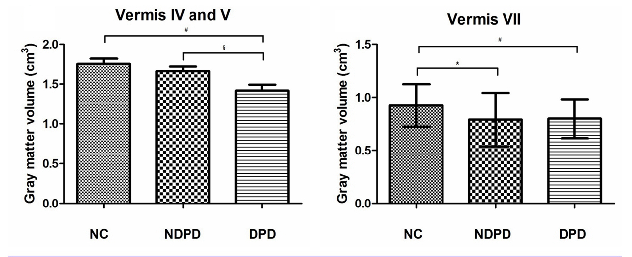
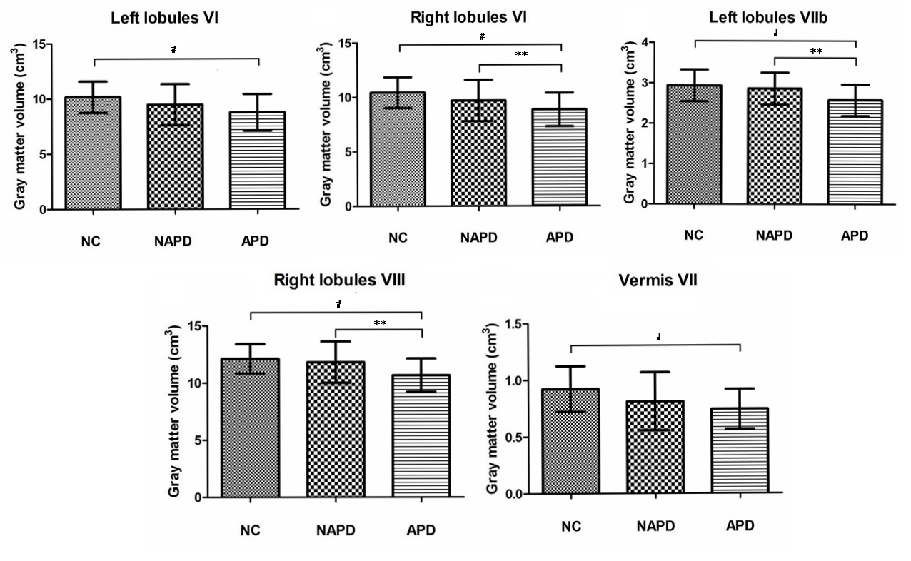
Results: When compared with NC, PDT group showed decreased GM volume in the right lobules IV, V, bilateral lobules VI and vermis VII. No difference was highlighted between PDAR and NC [figure1]. In addition, both DPD and NDPD groups exhibited reduced GM volume in vermis VII compared to NC group. Decreased GM volume was also found in the vermis IV, V of DPD group compared with NDPD and NC group respectively [figure2]. Furthermore, APD group showed decreased GM volume in the bilateral lobules VI, left lobules VIIb, right lobules VIII, and vermis VII compared to NC group. APD group also exhibited GM atrophy in the left lobules VIIb and the right lobules VI, VIII comparing with NAPD group. No difference was found between NAPD and NC [figure3].
Conclusions: Our study supports the possible role of the cerebellum in tremor-predominant subtype as well as the depressive and anxious symptoms in PD.
To cite this abstract in AMA style:
XX. Ma, CM. Li, W. Su, HB. Chen. Cerebellar atrophy in different motor and non-motor subtypes of Parkinson’s disease [abstract]. Mov Disord. 2018; 33 (suppl 2). https://www.mdsabstracts.org/abstract/cerebellar-atrophy-in-different-motor-and-non-motor-subtypes-of-parkinsons-disease/. Accessed January 22, 2026.« Back to 2018 International Congress
MDS Abstracts - https://www.mdsabstracts.org/abstract/cerebellar-atrophy-in-different-motor-and-non-motor-subtypes-of-parkinsons-disease/


