Session Information
Date: Thursday, June 8, 2017
Session Title: Parkinson's Disease: Neuroimaging And Neurophysiology
Session Time: 1:15pm-2:45pm
Location: Exhibit Hall C
Objective: To investigate resting state functional connectivity (rsFC) abnormalities in patients with Parkinson’s disease (PD), either with (PD-FOG) or without freezing of gait (PD no-FOG).
Background: Patients with PD develop several gait disturbances including FOG, a common symptom that deteriorates daily mobility leading to increased risk of falls and related injuries. Pathophysiological and neuroimaging studies demonstrated abnormalities in several brain areas involved in gait inhibition in PD [1-2]. In this study, we aimed to assess possible disturbances associated with intra-network and inter-network rsFC in patients with PD, PD-FOG and PD no-FOG.
Methods: Thirty-one PD patients, (15 PD-FOG and 16 PD no-FOG), and 16 healthy subjects (HS) underwent resting-state fMRI. The preprocessing steps included high pass filtering, spatial smoothening, motion correction, and normalization. Resting-state networks (RSNs) were further extracted to evaluate the within- and between-network rsFC using Melodic and FSLNets software packages. Group level differences were reported after correcting for multiple comparisons at p<0.05.
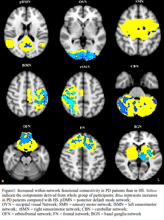
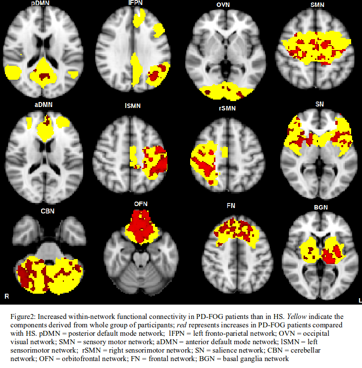
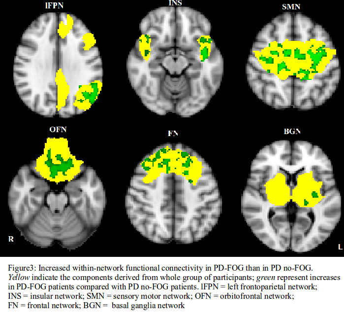
Results: PD patients, both PD-FOG and PD no-FOG groups, showed higher within-network rsFC than HS [figure1]. A larger number of RSNs was involved in PD-FOG, both when compared to HS [figure2] and to PD no-FOG [figure3]. PD no-FOG did not show increased rsFC in any of the RSNs relative to PD-FOG.
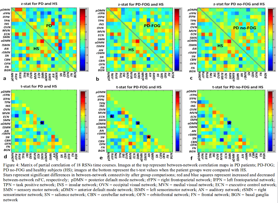
With respect to HS, the between-network rsFC among the anterior default mode network and sensory motor network decreased in PD as well as PD-FOG and PD no-FOG groups considered separately. Furthermore, PD-FOG showed increased rsFC between salience and auditory network as well as decreased rsFC between executive control network and right frontoparietal network compared with HS [figure4].
Conclusions: The within- and between-network findings clearly indicate a functional brain reorganization occurring in PD patients, with more severe abnormalities in PD-FOG compared with PD no-FOG. Decreased between-network rsFC between default mode network and sensory motor network in PD-FOG as well as in PD no-FOG may represent the functional substrate underlying motor and cognitive impairment in PD patients. Altered rsFC between salience and auditory network as well as between executive control network and right frontoparietal network may represent a specific pattern of rsFC in PD-FOG.
References: 1. Lewis, S. J., & Barker, R. A. (2009). A pathophysiological model of freezing of gait in Parkinson’s disease. Parkinsonism & related disorders, 15(5), 333-338.
2. Tessitore, Alessandro, et al. “Resting-state brain connectivity in patients with Parkinson’s disease and freezing of gait.” Parkinsonism & related disorders 18.6 (2012): 781-787.
To cite this abstract in AMA style:
K. Bharti, A. Suppa, N. Upadhyay, S. Pietracupa, G. Leodori, A. Zampogna, C. Gianni, N. Petsas, A. Berardelli, P. Pantano. Resting state functional connectivity in PD patients with and without Freezing of gait [abstract]. Mov Disord. 2017; 32 (suppl 2). https://www.mdsabstracts.org/abstract/resting-state-functional-connectivity-in-pd-patients-with-and-without-freezing-of-gait/. Accessed March 31, 2025.« Back to 2017 International Congress
MDS Abstracts - https://www.mdsabstracts.org/abstract/resting-state-functional-connectivity-in-pd-patients-with-and-without-freezing-of-gait/


