Session Information
Date: Wednesday, June 22, 2016
Session Title: Parkinson's disease: Neuroimaging and neurophysiology
Session Time: 12:00pm-1:30pm
Location: Exhibit Hall located in Hall B, Level 2
Objective: We aimed to improve our understanding of the neuropathology of excessive daytime sleepiness (EDS) in Parkinson’s disease (PD).
Background: EDS is a common non-motor symptoms in PD, but its neuropathology remains elusive due to limited studies and the inclusion of medicated patients. In this study, we investigated the underlying neuropathology associated with EDS in drug-naïve PD patients using a non-invasive neuroimaging technique.
Methods: A total of 76 PD patients in the early disease stages were recruited; 16 of them had EDS, while the remaining 60 did not. Resting state functional magnetic resonance imaging (rs-fMRI) was used to determine group differences (patients with EDS vs. patients without EDS) in spontaneous neural activity indicated by regional homogeneity (ReHo). Additionally, functional connectivity (FC) of the regions showing group differences in ReHo with the entire brain was performed.
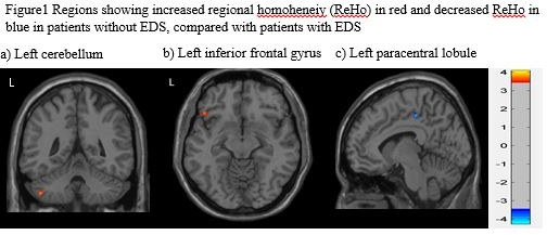
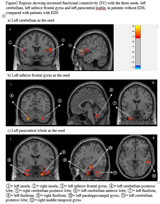
Results: No group difference in demographic and clinical variables was noted, except that patients with EDS showed more severe postural instability gait difficulty (PIGD, p= 0.035) and rapid eye movement sleep behavior disorder (RBD, p= 0.006), compared to patients without EDS. ReHo analysis controlling for gray matter volume, age, gender, general cognition, depression, PIGD, and RBD showed decreased ReHo in the left cerebellum and inferior frontal gyrus, but increased ReHo in the left paracentral lobule in PD-EDS patients, compared with patients without EDS. Further, EDS severity was inversely correlated with ReHo signals in the left cerebellum posterior lobe (r= -0.33, p<0.01) and positively correlated with ReHo signals in the left paracentral lobule (r= 0.24, p < 0.05), while the correlation between EDS severity and ReHo signals in the left inferior frontal gyrus approached significance (r= -0.21, p= 0.069). FC analysis controlling for the same variables as in the analysis of ReHo revealed that the three regions showing ReHo differences had decreased FC with regions in the frontal, temporal, insular and limbic lobes and cerebellum in PDs with EDS. 

Conclusions: While decreases in ReHo and FC were found, increases in ReHo were also noted, implying both neural downregulation and compensatory mechanisms in early PD patients with EDS. Longitudinal studies are warranted to clarify the long-term impact of EDS in PD.
To cite this abstract in AMA style:
M.C. Wen, S.Y.E. Ng, H.S.E. Heng, Y. Chao, L.L. Chan, E.K. Tan, L.C.S. Tan. Neural substrates of excessive daytime sleepiness in early drug naïve Parkinson’s disease: A resting state functional MRI study [abstract]. Mov Disord. 2016; 31 (suppl 2). https://www.mdsabstracts.org/abstract/neural-substrates-of-excessive-daytime-sleepiness-in-early-drug-nave-parkinsons-disease-a-resting-state-functional-mri-study/. Accessed April 2, 2025.« Back to 2016 International Congress
MDS Abstracts - https://www.mdsabstracts.org/abstract/neural-substrates-of-excessive-daytime-sleepiness-in-early-drug-nave-parkinsons-disease-a-resting-state-functional-mri-study/
