Session Information
Date: Wednesday, June 7, 2017
Session Title: Parkinson's Disease: Cognition
Session Time: 1:15pm-2:45pm
Location: Exhibit Hall C
Objective: To explore the association of acyl-ghrelin (AG) levels with cognitive impairment (CI) in PD
Background: Unintended weight loss (UWL) in PD is associated with CI. AG is produced in response to weight loss and stimulates appetite and weight gain. Levels are paradoxically low in people with PD and UWL[1]. AG can improve learning and memory in animal studies through promotion of long-term potentiation and neurogenesis. It is anti-inflammatory and stabilises mitochondria, preventing neuronal apoptosis. Thus, it has been shown to be neuroprotective in animal models of PD[2]. AG indirectly stimulates IGF-1; also proposed to be neuroprotective in PD. AG may therefore provide a mechanistic neurohumoral link between UWL and CI in PD. No studies have explored the association of ghrelin with CI in PD to date.
Methods: 55 adults aged 60-85 were recruited; healthy controls (HC) (n=20), PD without CI (PD-NC)(n=19) and PD with CI (PD-CI)(n=16). The PD-CI group included PDD and PD-MCI. All had a Montreal Cognitive Assessment of ≤25/30. Participants with UWL, obesity, BMI <18 or >30, diabetes, gastrointestinal disease, smoking, deep brain stimulation or non-selective anticholinergic medication were excluded. Participants were tested fasted and off PD medication. Blood was drawn at baseline, 5, 15, 30, 60, 120 and 180 minutes following a standard breakfast. AG samples were treated with 4-(2-Aminoethyl)-benzenesulfonyl fluoreide and analysed using a multiplex assay. Total ghrelin (TG), IGF-1 and GH were analysed by ELISA. At 180 minutes participants received an ad libitum meal. Area under the curve (AUC) was calculated for each analyte using the trapezoidal method and AUC compared using analysis of variance. Post-hoc comparisons were conducted by Scheffe’s test.
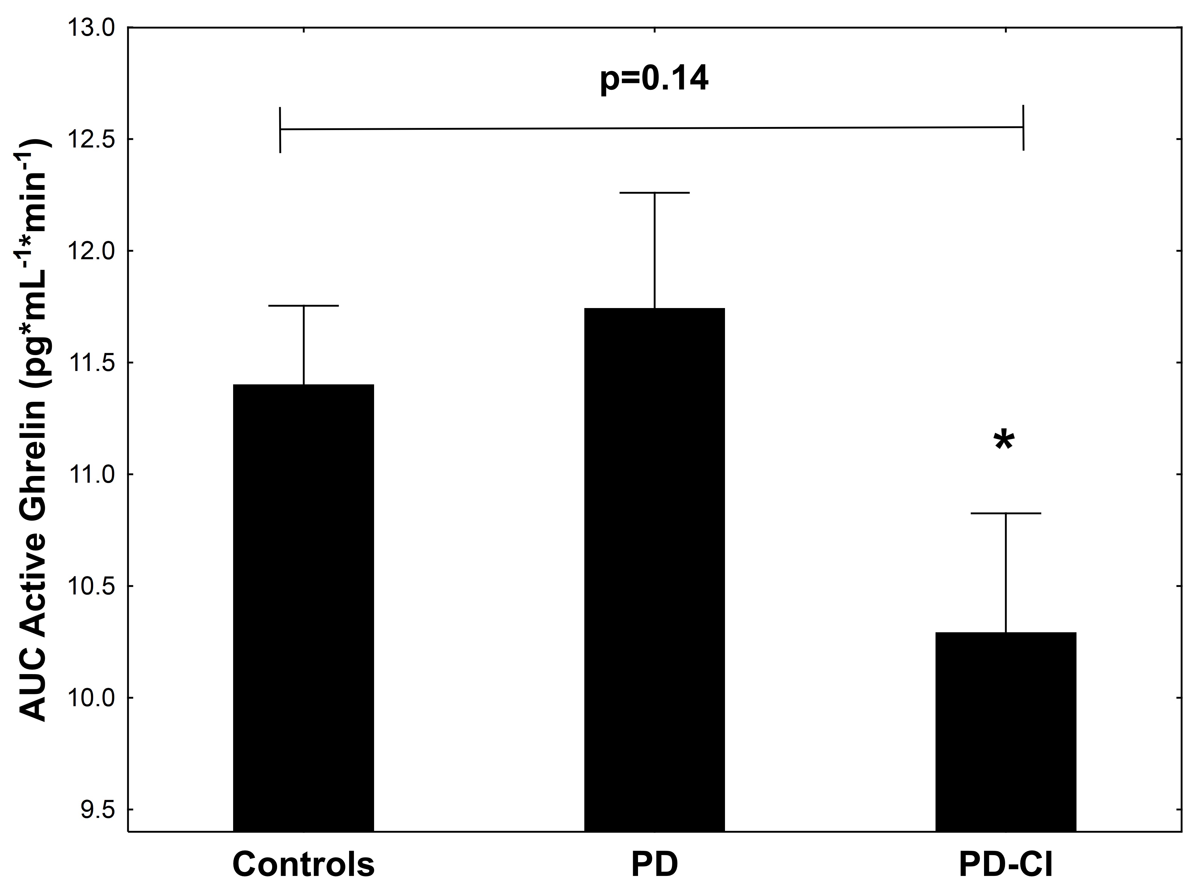
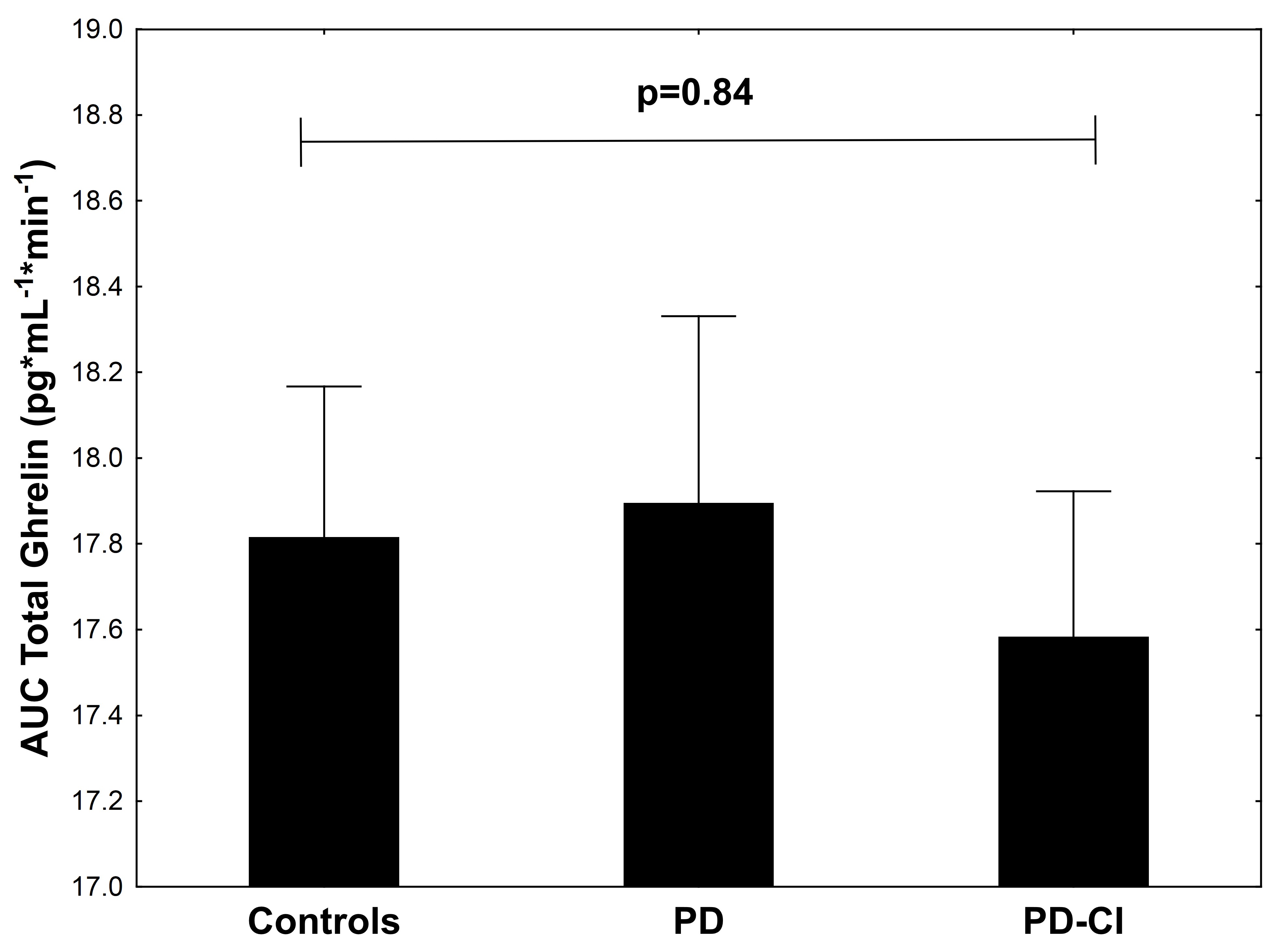
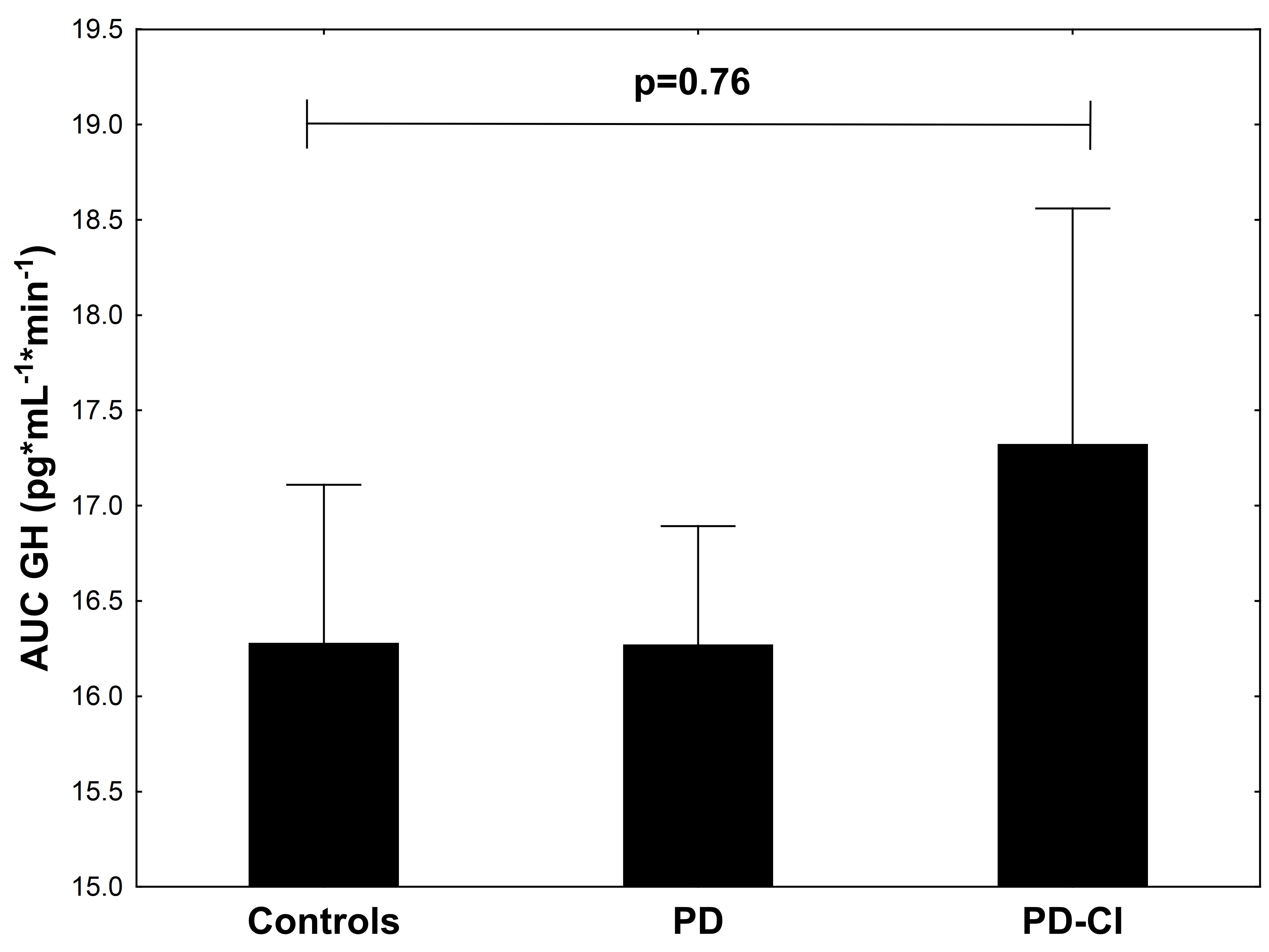
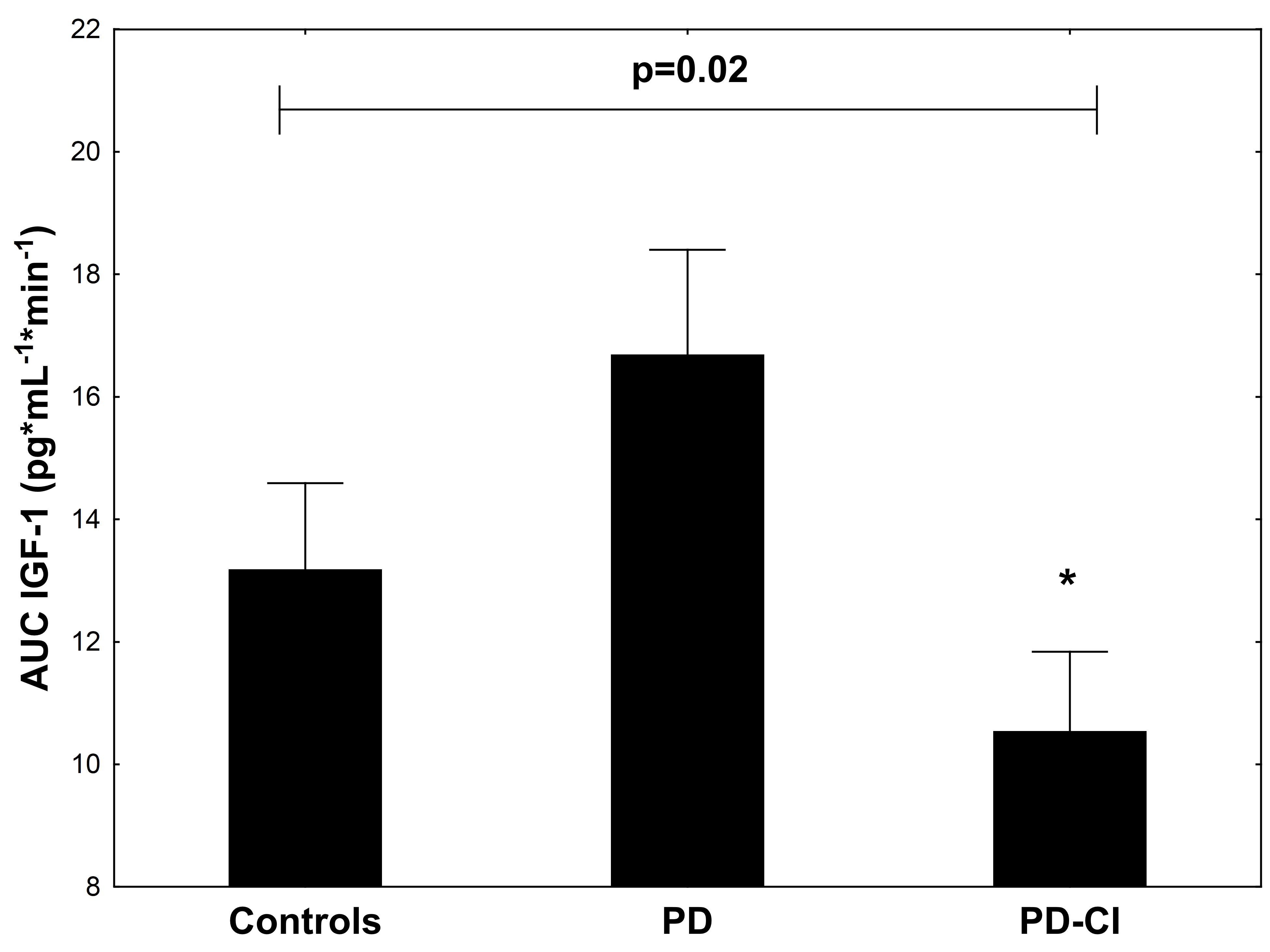
Results: There were no significant differences between groups for age, gender or disease duration. AUC for AG showed a trend towards lower levels in the PD-CI group (p=0.14), which had significantly lower levels compared to the PD group (p<0.05)[Figure 1], There was no significant difference between groups for TG (p=0.84)[Figure 2]or GH (p= 0.76)[Figure 3]. There was a significant difference between groups for IGF-1 (p=0.02)[Figure 4], with lower levels seen in PD-CI(p<0.05).
Conclusions: IGF-1 levels were significantly different between HC, PD and PD-CI. AG levels were significantly lower in PD-CI than PD. AG and IGF-1 should be investigated as possible biomarkers for CI in PD with longitudinal studies.
References: 1. Fiszer, U., et al., Leptin and ghrelin concentrations and weight loss in Parkinson’s disease. Acta Neurologica Scandinavica, 2010. 121(4): p. 230-236.
2. Moon, M., et al., Neuroprotective effect of ghrelin in the 1-methyl-4-phenyl-1, 2, 3, 6-tetrahydropyridine mouse model of Parkinson’s disease by blocking microglial activation. Neurotoxicity research, 2009. 15(4): p. 332-347.
To cite this abstract in AMA style:
F. Johnston, M. Siervo, A. Hornsby, J. Davies, D. Burn. Ghrelin and the IGF-1 axis in cognitive impairment in PD [abstract]. Mov Disord. 2017; 32 (suppl 2). https://www.mdsabstracts.org/abstract/ghrelin-and-the-igf-1-axis-in-cognitive-impairment-in-pd/. Accessed February 18, 2026.« Back to 2017 International Congress
MDS Abstracts - https://www.mdsabstracts.org/abstract/ghrelin-and-the-igf-1-axis-in-cognitive-impairment-in-pd/


