Session Information
Date: Monday, June 20, 2016
Session Title: Surgical therapy: Other movement disorders
Session Time: 12:30pm-2:00pm
Location: Exhibit Hall located in Hall B, Level 2
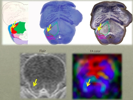
Objective: We investigate DTI and Fractional Anisotropy maps both in vivo and in post-mortem MR studies. In situ imaging findings were correlated with those obtained from actual histological sections from the same specimen.
Background: The Pedunculopontine Nucleus (PPN) is a brainstem nucleus involved in the control of muscle tone and locomotion. It has been proposed as target for deep brain stimulation (DBS) in patients with gait disorders. Although studies have suggested that diffusion tensor imaging (DTI) may help guiding electrode placement in the PPN, none have provided a direct histological correlation to date.
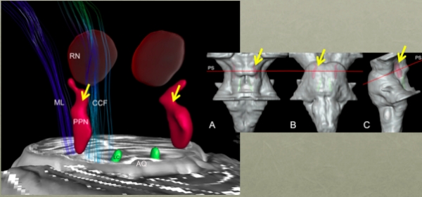
Methods: A post-mortem brain was scanned in a clinical 3T MR system before procurement (in cranio). The brain was processed with a special method ideally suited for cytoarchitectonic analyses. Nine in vivo control volunteers were scanned using the same MRI protocol. Images from the volunteers were compared to those obtained in the post-mortem study. FA values were obtained from PPN, inferior colliculus (IC), cerebellar crossing fibers (CCF) and medial lemniscus (ML) using the histological data and atlas information.
Results: FA values in the PPN were significantly lower than in the surrounding white matter region and higher than in gray matter areas. Nissl-stained histologic series the PPN extended for more than 10mm in rostro-caudal direction and it was closely attached to the lateral parabrachial nucleus (PBL). 
 Our DTI analyses and the spatial correlation with histological findings proposed a location for PPN that matched the position assigned to this nucleus by literature.
Conclusions: Coregistration of neuroimaging and cytoarchitectonic features can add value to help establishing functional architectonics of the PPN and facilitate neurosurgical targeting of this extended nucleus.
To cite this abstract in AMA style:
A.T.D.L. Alho, C. Hamani, E.J.L. Alho, R.E. da Silva, G.A.B. dos Santos, R.C. Neves, C.M.M. Araujo, G. Magalhães, L.T. Grinberg, H. Heinsen, E.T. Fonoff, E. Amaro-Junior. Magnetic resonance diffusion tensor imaging for the pedunculopontine nucleus: Correlation between neuroimaging and cytoarchitectonic features [abstract]. Mov Disord. 2016; 31 (suppl 2). https://www.mdsabstracts.org/abstract/magnetic-resonance-diffusion-tensor-imaging-for-the-pedunculopontine-nucleus-correlation-between-neuroimaging-and-cytoarchitectonic-features/. Accessed December 26, 2025.« Back to 2016 International Congress
MDS Abstracts - https://www.mdsabstracts.org/abstract/magnetic-resonance-diffusion-tensor-imaging-for-the-pedunculopontine-nucleus-correlation-between-neuroimaging-and-cytoarchitectonic-features/
