Session Information
Date: Wednesday, June 22, 2016
Session Title: Neuroimaging (non-PD)
Session Time: 12:00pm-1:30pm
Location: Exhibit Hall located in Hall B, Level 2
Objective: To explore the role of GABA in the pathophysiology of ET and DT by using MR spectroscopy.
Background: The mechanisms of tremor in ET and dystonic tremor (DT) are still unclear. Previous evidence lead to the hypothesis of disturbances of GABA in the cerebellum in ET whereas it is unknown in DT.
Methods: 12 ET, 11 DT patients and 10 controls were scanned on a 3-Tesla GE MRI scanner with a 32-channel coil. High-resolution T1-weighted images obtained with a magnetization-prepared gradient-echo (MPRAGE) sequence were used for localization and tissue composition estimation. Two voxels of interest (30x30x20 mm) were centered over the right cerebellar hemisphere (CB) and the mid frontal lobe (MF), as control. We measured metabolites using the point-resolved spectroscopy (PRESS) pulse sequence and the J-edited method (TR/TE 1500/68 ms, 768 acquisitions, ∼20 min scanning time). GABA peak, including macromolecules, (GABA+) at 3 ppm and Glx (glutamate and glutamine) at 3.8 ppm were fitted using in-house software written in IDL by JWvdV. Both peaks were referenced to Creatine (Cre) as it has steady concentration in the brain. Using FSL and AFNI software libraries, we calculated the percentage of CSF, grey (GM) and white matter (WM) in each voxel.
Results: The demographic data are shown in table 1.
| Character | ET (N=12) | DT (N=11) | Controls (N=10) | P value |
| Age | 58 ± 9.5 | 59.7 ± 13.2 | 60.6 ± 8.5 | 0.84 |
| Female (%) | 5 (42%) | 5 (45%) | 5 (50%) | 0.93 |
| Tremor duration | 29.9 ± 16.3 | 27.8 ± 18.6 | 0.61 | |
| Tremor onset | 28.1 ± 20.5 | 32 ± 18.6 | 0.76 | |
| Dystonia duration | 17 ± 16.6 | |||
| Dystonia onset | 42.4 ± 16.8 | |||
| TETRAS ADL score | 21.8 ± 4.2 | 11.3 ± 7.3 | < 0.01 | |
| TETRAS performance | 17.7 ± 5.5 | 12.7 ± 6.3 | 0.05 | |
| TETRAS total score | 39.5 ± 8.4 | 24.5 ± 11.6 | < 0.01 |
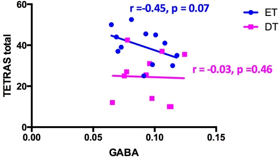
 Percentages of GM and WM in the CB voxel were significantly different among groups while the voxel location was not different. There was a trend for a negative correlation between TETRAS score and GABA+/Cre in the CB only in the ET (r=-0.45,p=0.07) but not with DT (r=-0.03,p=0.46).
  However, there were no correlations between GABA+/Cre with disease duration and tissue composition.
Conclusions: Our study found no differences in GABA levels in cerebellum between ET, DT and controls using MR spectroscopy. This could be related to the voxel and sample size, or could indicate no changes in overall concentration of GABA in CB in ET and DT. However, a trend of negative correlation between GABA composition in the CB and disease severity scale in ET might support the role of cerebellar GABAergic dysfunction in ET but not DT. Additionally, further morphometric studies of the whole CB in both ET and DT are warranted.
To cite this abstract in AMA style:
P. Panyakaew, H.J. Cho, S. Horovitz, M. Hallett. Cerebellar GABA in essential tremor and dystonic tremor: A MR spectroscopy study [abstract]. Mov Disord. 2016; 31 (suppl 2). https://www.mdsabstracts.org/abstract/cerebellar-gaba-in-essential-tremor-and-dystonic-tremor-a-mr-spectroscopy-study/. Accessed February 20, 2026.« Back to 2016 International Congress
MDS Abstracts - https://www.mdsabstracts.org/abstract/cerebellar-gaba-in-essential-tremor-and-dystonic-tremor-a-mr-spectroscopy-study/
