Session Information
Date: Monday, June 5, 2017
Session Title: Parkinsonism, MSA, PSP (Secondary and Parkinsonism-Plus)
Session Time: 1:45pm-3:15pm
Location: Exhibit Hall C
Objective: To do (1) H NMR study in a group of Parkinson’s disease patients, disease controls, viz. multiple system atrophy (MSA) and progressive supra nuclear palsy (PSP) to explore disease-specific metabolic signatures.
Background: Parkinson’s disease (PD) is a debilitating movement disorder resulting from a progressive degeneration of the nigrostriatal dopaminergic pathway and depletion of neurotransmitter dopamine in the striatum. To date, available data have not yet revealed one reliable biomarker to detect early neurodegeneration in PD and to detect and monitor effects of drug candidates on the disease process.
Methods: UK PDS Brain Bank criteria for the diagnosis of PD were applied to select the patients. Serum from blood was frozen at −80°C until NMR analysis was performed. NMR experiments were conducted on Bruker Avance DRX 800 MHz spectrometer equipped with a cryoprobe with a Z-shielded gradient (Bruker Biospin, Karlsruhe, Germany). One-dimensional (1)H NMR measurements were obtained using Carr–Purcell–Meiboom–Gill (CPMG) sequence with water suppression by pre-saturation at 300 K.
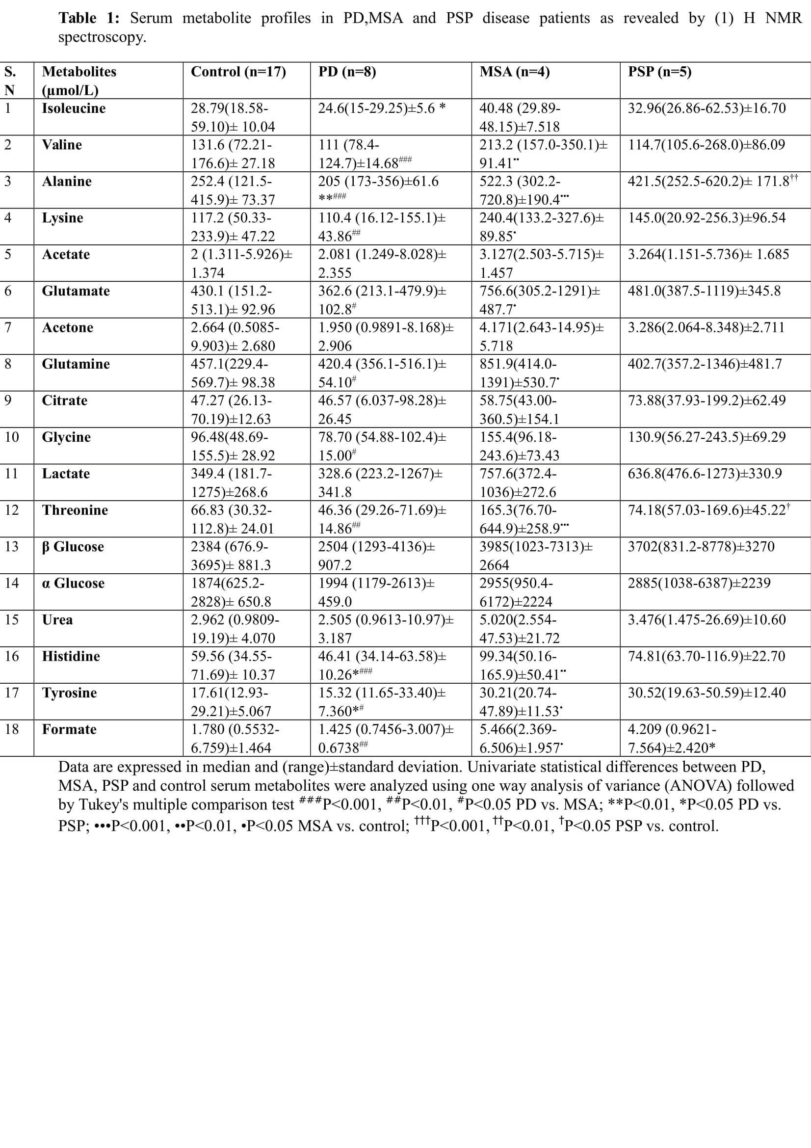
Results: As demonstrated by (1) H NMR spectroscopy, serum Valine, Alanine, Histidine Lysine, Threonine, Formate and Glutamate, Glutamine, Glycine, Tyrosine concentrations were significantly different in the patients of PD and MSA. Serum concentrations of Isoleucine, Histidine, Tyrosine, Formate and Alanine were significantly different in MSA and PSP. Whereas, the serum concentrations of Alanine, Threonine, Valine, Histidine, Lysine, Glutamate, Glutamine, Tyrosine, Formate were significantly different in MSA with respect to healthy controls. As far as PSP vs. Control concerned, Alanine and Threonine were significantly different with respect to controls.
Conclusions: Observations include increased glutamate levels in MSA, which consolidates the hypothesis of excitatory amino acid glutamate involvement in neurodegeneration. We also observed changes in other amino acid levels. Formate is increased in both MSA and PSP. It is responsible for both metabolic acidosis and disrupting mitochondrial electron transport and energy production by inhibiting cytochrome oxidase activity.
To cite this abstract in AMA style:
G.N. Babu, M. Gupta, V.K. Paliwal, S. Singh, R. Roy. Metabolomics study in a group of Parkinson’s disease patients from Northern India [abstract]. Mov Disord. 2017; 32 (suppl 2). https://www.mdsabstracts.org/abstract/metabolomics-study-in-a-group-of-parkinsons-disease-patients-from-northern-india/. Accessed December 18, 2025.« Back to 2017 International Congress
MDS Abstracts - https://www.mdsabstracts.org/abstract/metabolomics-study-in-a-group-of-parkinsons-disease-patients-from-northern-india/

