Session Information
Date: Wednesday, June 22, 2016
Session Title: Neuroimaging (non-PD)
Session Time: 12:00pm-1:30pm
Location: Exhibit Hall located in Hall B, Level 2
Objective: Use modified histological techniques and a computational registration pipeline to delineate accurately post-operative 3D basal ganglia anatomy in DBS implants.
Background: Stereotaxy is based on the precise image-guided spatial localization of targets within the human brain. Unfortunately, it is still not possible to reliably delineate all the sub-cortical structures using MRI. Although several maps have been selected as a basis for correlating imaging results with the anatomical locations of these structures, technical limitations interfere in a point-to-point correlation between imaging and individual anatomy.
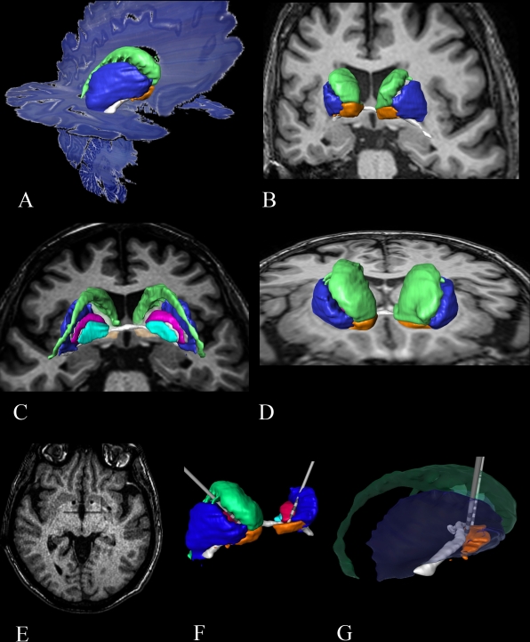
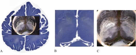
Methods: A post-mortem 66 year-old brain was scanned in a 3.0T MRI. The brain was fixed in formaline, embedded in celloidin and serially sectioned in the axial plane with a thickness of 400µm. The slices were stained with gallocyanin and mounted as described by Heinsen et al., 2000. Our registration pipeline included blockface and histology segmentation, followed by symmetrical 2D affine registration based on robust statistics and 2D and 3D non-linear ANTS SyN registration. Details on the pipeline were presented in Alegro et al., 2015. The outlines of basal ganglia structures were segmented in high resolution dark field images and saved as binary masks. The 2D and 3D transformation maps were applied to these masks. Subsequently, the post-mortem in cranio MRI and the cytoarchitectonic maps were registered onto a post-operative patient’s MRI. This patient is a 62 years old woman suffering from refractory OCD, submitted to DBS of the ventral striatum.
Results: Our previous results suggest good localization and highly similar geometry when comparing the registered histological volume and the MRI. Cytoarchitetonic segmentations of basal ganglia structures were deformed using the maps computed during the pipeline execution, producing a coherently registered 3D impression of these nuclei. The dark field high resolution maps were transferred to post-operative MRI rendering a clear 3D localization of the trajectory and localization of the leads within the basal ganglia. 

Conclusions: Modified histological techniques to prevent deformations and enhance segmentation together with a computational registration pipeline can register highly accurate cytoarchitetonic maps to post-operative patient’s MRI.
To cite this abstract in AMA style:
E.J.L. Alho, M. Alegro, R.L. Deus, R.P. Reis, L.T. Grinberg, J.F. Pereira Jr, L. Zöllei, E. Amaro Jr., H. Heinsen, E.T. Fonoff. Three-dimensional cytoarchitecture of the basal ganglia: Clinical applications of a computational pipeline for histology to MRI registration [abstract]. Mov Disord. 2016; 31 (suppl 2). https://www.mdsabstracts.org/abstract/three-dimensional-cytoarchitecture-of-the-basal-ganglia-clinical-applications-of-a-computational-pipeline-for-histology-to-mri-registration/. Accessed December 19, 2025.« Back to 2016 International Congress
MDS Abstracts - https://www.mdsabstracts.org/abstract/three-dimensional-cytoarchitecture-of-the-basal-ganglia-clinical-applications-of-a-computational-pipeline-for-histology-to-mri-registration/
